Friday, October 29, 2010
Monday, September 20, 2010
Saturday, September 4, 2010
sinogram+sub periosteal abscess
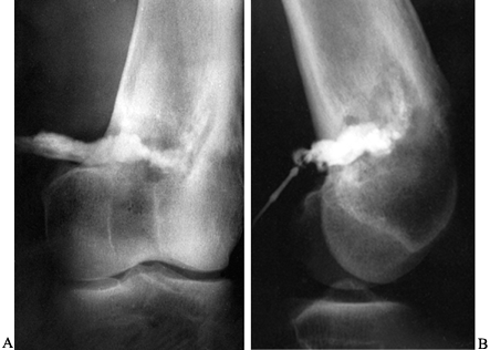
Lateral radiograph (A) of a 15-year-old boy shows a subperiosteal abscess that was aspirated and injected with contrast material. Anteroposterior (B) and lateral (C) radiographs of a 9-year-old child show contrast material in the subperiosteal area and within the medullary cavity after pus had been aspirated. The contrast material demonstrates the extent of the abscess.
sinogram+osteomylitis
sinogram
What is sinogram?
It is a special X-ray procedure that is done with contrast dye to visualize any abnormal opening (sinus) in the body. The contrast is injected via a rubber catheter. Serial x-ray pictures are taken to show the extension of the fistula.
What kind of contrast medium used in this procedure?
A low osmolar contrast medium, LOCM 150.
How is it done?
Technique :
- A prelim film is taken to exclude the presence of radio-opaque foreign body.
- A fine catheter is then inserted into the orifice of the sinus.
- After a gauze pad has been firmly placed over the orifice to discourage reflux, the contrast medium is injected under fluoroscopic control.
- Spot films are taken as required including tangential views.
Below are sequence of films taken to investigate a sinus at right mid thigh.
1. Prelim : shows intramedullary fixation of right femur fracture. Site of fistula is located at mid thigh.
2. Right thigh AP : Dye is injected. There is a focal collection of contrast seen.
3. Lateral view : shows focal collection of contrast.
4. Right lower thigh AP : shows seepage of contrast seen into the intramuscular layers of the lateral aspect of the right thigh.
5. Right upper thigh AP : shows seepage of contrast seen into the intramuscular layers of the lateral aspect of the right thigh up to the level of hip joint and distally to the level of distal femur (just above the femoral condyles).
How is it reported?
This is a sample report of a sinogram case.NAME : ?
I/C : ?
SINOGRAM (01.04.2010)
Procedures:
Patient wound is cleaned. Sinus identified.
25 ml undiluted omipaque injected using 8F nasogastric tube.
Serial x-rays are taken.
Findings:
There is flow of contrast from the sinus into a focal collection measuring 3.5×4.5cm.
Seepage of contrast seen into the intramuscular layers of the lateral aspect of the right thigh.
Superiorly the contrast extends to the level of hip joint and distally to the level of distal femur (just above the femoral condyles).
There is no connection to the knee or hip joint.
CONCLUSION
No evidence of intra-articular extension of the right thigh abscess.
Radiologist 01.04.2010
Reference :
- A Guide to Radiological Procedures Stephen Chapman.


Saturday, July 31, 2010
Plantar Fasciitis
-The sagittal STIR (edema sensitive) MR image shows a thickened plantar fascia with edema. Contrast this with the thin fascia seen on the normal sagittal view.-The coronal PD (proton density) MR image also shows thickening and increased signal in the fascia.
Discussion
When patient's present to a doctor complaining of heel pain, plantar fasciitis is the most common cause. It is caused by repetitive microtrauma and is common in runners.
http://eradiology.bidmc.harvard.edu/Classics/item.aspx?section=Emergency+Radiology&labelpk=33f0adab-853f-4010-a0e2-0756b3f1eac5&pk=d9066408-e848-453f-a2dd-ecbe3b9786d7
Elbow: Biceps Tendon Rupture on MRI Scan
Findings-The T2 MR image with fat saturation shows a ruptured distal biceps brachii tendon.

-The tendon is wavy and there is edema in the cubital fossa.
Discussion
Biceps tendon ruptures usually occur due to trauma from a forceful flexion of the elbow. Visualization the ends of the tendons is crucial for preoperative planning.
http://eradiology.bidmc.harvard.edu/Classics/item.aspx?section=Emergency+Radiology&labelpk=33f0adab-853f-4010-a0e2-0756b3f1eac5&pk=468a2719-c9b7-4f6d-9c9e-cc181a5b059b
Tuesday, July 27, 2010
Calcaneal fractures. Lateral ankle radiograph of 56-year-old woman with diabetes reveals avulsion fracture of calcaneal tuberosity.http://www.ajronline.org/cgi/content/full/182/1/147/FIG10
Calcaneal fractures. Lateral ankle radiograph of 38-year-old woman who presented with ankle pain shows curvilinear sclerosis (arrows) exiting into superior aspect of calcaneus, consistent with stress fracture. Note subtle density in Kager's fat pad (asterisk).http://www.ajronline.org/cgi/content/full/182/1/147/FIG9
32-year-old woman with Haglund's disease. Lateral ankle radiograph shows thickening of Achilles tendon at insertion (asterisk), retrocalcaneal bursitis (curved arrowhead), and retro-Achilles bursitis (double arrows). Triad of findings is consistent with Haglund's disease.http://www.ajronline.org/cgi/content/full/182/1/147/FIG7
Abnormalities of Achilles tendon. Lateral ankle radiograph of 48-year-old man shows ossified Achilles tendon caused by previous tear.http://www.ajronline.org/cgi/content/full/182/1/147/FIG5
Abnormalities of Achilles tendon. Sagittal spin-echo T1-weighted image of 42-year-old man shows large area of intermediate signal in expected location of Achilles tendon, flanked superiorly and inferiorly by irregular ends of ruptured and retracted Achilles tendon.http://www.ajronline.org/cgi/content/full/182/1/147/FIG4
Abnormalities of Achilles tendon. Sagittal fat-suppressed fast spin-echo T2-weighted image of 26-year-old man shows focal hyperintensity in thickened Achilles tendon, corresponding to partial tear (arrowhead) of Achilles tendon, and edema (arrow) in Kager's fat pad.http://www.ajronline.org/cgi/content/full/182/1/147/FIG3
Kager's fat pad
Normal anatomy of Kager's fat pad. Lateral radiograph (A) and illustration (B) of ankle show triangular radiolucency (A) and structure of Kager's fat pad. On radiograph, anterior border (arrowheads, A) is posterior aspect of flexor hallucis longus muscle and tendon, posterior border (asterisk, A) is Achilles tendon, and floor (arrow, A) is superior surface of calcaneus.http://www.ajronline.org/cgi/content/full/182/1/147/FIG2
Normal anatomy of Kager's fat pad. Lateral radiograph (A) and illustration (B) of ankle show triangular radiolucency (A) and structure of Kager's fat pad. On radiograph, anterior border (arrowheads, A) is posterior aspect of flexor hallucis longus muscle and tendon, posterior border (asterisk, A) is Achilles tendon, and floor (arrow, A) is superior surface of calcaneus.http://www.ajronline.org/cgi/content/full/182/1/147
FindingsThe axial MRI of the shoulder shows increased T2 signal in the long head of the biceps tendon in the bicipital groove compatible with tendinopathy.
http://eradiology.bidmc.harvard.edu/Classics/item.aspx?section=Emergency+Radiology&labelpk=33f0adab-853f-4010-a0e2-0756b3f1eac5&pk=96e27e50-c28e-4dc9-a0e1-2f47fb3e4998
Findings1-The Achilles tendon is the largest tendon in the body. It is formed from the conjoined tendons of the gastrocnemius and soleus muscles. It inserts on the posterior calcaneus.
2-The Achilles tendon contributes to plantar flexion of the foot.
3-Tears occur at the vulnerable zone of avascularity, 2-6 centimeters above the calcaneal insertion.
4-This patient’s plain film shows edema in the Kagers fat pad suggesting Achilles tendon pathology. Kager's fat pad normally presents as a lucent triangle above the superior calcaneus. The region shows abnormal soft tissue density in this patient.
5-The T2 weighted Sagittal MRI shows discontinuity of the Achilles tendon with fluid at the myotendinous junction.
http://eradiology.bidmc.harvard.edu/Classics/item.aspx?section=Emergency+Radiology&labelpk=33f0adab-853f-4010-a0e2-0756b3f1eac5&pk=37363e43-d4af-4d51-b254-632023ada1e6
Friday, May 28, 2010
The axial image demonstrates increased signal (arrow) within fat interposed between the lateral aspect of the patellar tendon and the lateral trochlear ridge. Mild lateral patellar subluxation is present as indicated by the off-midline positioning of the patellar tendon (arrowhead).
The sagittal image demonstrates increased signal (arrow) within the fat below the caudal margin of the patella as well as moderate patella alta (arrowhead). Note the high riding appearance of the patella.
Three normal anterior knee fat pads are present at the knee; the quadriceps (anterior suprapatellar) (red), the prefemoral (posterior suprapatellar or supratrochelar) (blue), and Hoffa (infrapatellar) (yellow) fat pads.The infrapatellar fat pad is bordered by the inferior pole of the patella superiorly, the joint capsule and patellar tendon anteriorly, the proximal tibia and deep infrapatellar bursa inferiorly, and the synovium-lined joint cavity posteriorly. Thus, it is intracapsular but extrasynovial. It is tethered to the intercondylar notch superiorly by the infrapatellar synovial fold or infrapatellar plica. It also is attached directly to the anterior horns of the menisci inferiorly and to the periosteum of the tibia.
On MRI, a focal area of high signal or edema on STIR or T2 weighted sequences is present at the inferolateral aspect of the patellofemoral joint, specifically the lateral portion of the infrapatellar fat pad (D,E). Specifically, this is just below the inferior margin of the patella and anterior to the lateral trochlear ridge. A focal lobulated mass with signal characteristics of fluid or cystic change can sometimes be present in the lateral soft tissues of the knee between the lateral femoral condyle and the lateral retinaculum. Associated findings of lateral patellar subluxation (F) and/or patella alta are frequently present.
http://www.radsource.us/clinic/0809
Tuesday, May 25, 2010
fat-suppressed proton density-weighted sagittal images in a basketball player who sustained an acute injury reveal marked edema about the patellar tendon with a fluid filled gap (arrows) at the central tendon, compatible with a complete rupture. The retracted proximal and distal ends (arrowheads) are markedly thickened, indicating severe tendinosis. The vast majority of patellar tendon ruptures occur in patients with pre-existing patellar tendinosis.
A proton density-weighted axial view demonstrates the normal semilunar appearance of the patellar tendon, with a convex anterior border (arrow) and well-defined posterior rim.
T1-weighted sagittal image in a patient with a normal patellar tendon reveals a homogeneously low signal intensity appearance to the tendon (arrows), which appears symmetrical in appearance throughout its course.http://www.radsource.us/clinic/0612