 Left testicular venogram. This image shows a left testicular varicocele before embolization. Note: radiographs of varicoceles should be avoided to restrict radiation exposure.
http://emedicine.medscape.com/article/382288-imaging


 Left testicular venogram. This image shows a left testicular varicocele before embolization. Note: radiographs of varicoceles should be avoided to restrict radiation exposure.
 Sonogram depicting a combination of an intratesticular and extratesticular right-sided varicocele.
 Transverse ultrasonography scan through the left testicle. This image shows several cystic structures (2.4-4 mm in diameter) behind the testis. Color flow Doppler image demonstrates flow within these cystic structures (not shown).
 Line diagram of a typical bidirectional Doppler recording of a stop-type varicocele (red) in which flow is stopped by a competent valve in the pampiniform plexus above the level of communicating veins. The shunt-type of varicocele is depicted in blue. Because the valves in the pampiniform plexus contiguous to the testicular veins are incompetent or absent, venous flow is shunted via communicating veins into orthograde draining veins; these are shown to represent deferential and cremasteric veins at contrast venography.
 Upper image: Longitudinal sonogram through the pampiniform plexus of the left testis. The image shows several anechoic tubes. Lower image: The application of color Doppler imaging in the same patient shows bidirectional flow within the anechoic tubes.
 Longitudinal sonogram through the left testicle. This image shows several large anechoic tubes (2.4-6 mm in diameter) lying behind the upper and middle poles of the testicle. The diameter of these tubes increased by 1.5-2 mm with a Valsalva maneuver (not shown). T = testicle; v = varicocele.

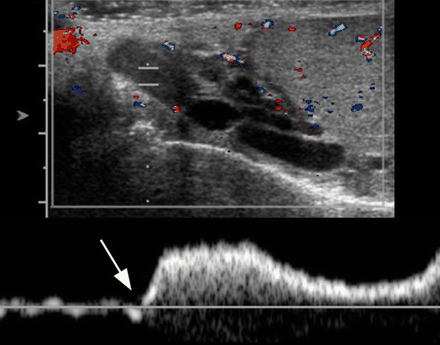 Left varicocele in a 15-year-old boy. (a) Longitudinal US scan of the left hemiscrotum shows multiple anechoic structures (arrows) in the supratesticular region and extending behind the upper pole of the testis (T). (b) Color Doppler image shows that the anechoic structures are vascular. (c) Pulsed-wave Doppler image shows a venous waveform with increased flow during the Valsalva maneuver (arrow).


