Best Bath Store
All Natural Blend of 14 Herbs Gentle Cleanse- $20 Off Colonix Now! Use Promo Code 20J
Amberen - Breakthrough Menopause Treatment
728x90
Custom Search
Showing posts with label epididymitis. Show all posts

Friday, June 11, 2010

Acute epididymitis in a 9-year-old boy with scrotal pain and redness. Longitudinal US scan shows that the epididymal head and body (arrows) are enlarged and hypoechoic relative to the normal testis (T). Wall thickening (*) and reactive hydrocele (h) are also seen. Power Doppler imaging showed increased perfusion of the epididymis.

http://radiographics.rsna.org/content/25/5/1197.figures-only
A
B
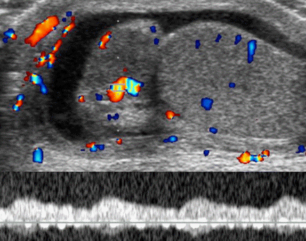
Clinically proved epididymitis in an 11-year-old boy. (a) Longitudinal US scan of the right hemiscrotum shows an enlarged hypoechoic epididymal head (E), reactive hydrocele (h), and thickening of the scrotal wall (*). m = mediastinum. (b) Color and pulsed-wave Doppler image shows increased vascularity in the epididymal head with a low-flow, low-resistance waveform pattern.
Sonographically the acute epididymitis usually shows enlargement of the epididymal head with decreased echogenicity secondary to edema. A reactive hydrocele may be present. Color Doppler findings include an increased amount of flow in and around the epididymis. If an abscess has formed, complex cystic areas may be identified in the epididymis.


http://www.google.com.eg/imgres?imgurl=http://www.gehealthcare.com/usen/ultrasound/images/cme_scrotal5_500.jpg&imgrefurl=http://www.gehealthcare.com/usen/ultrasound/products/cme_scrotal.html&usg=__pRNzx5ouiF10cwNwsoFkjfJSRXA=&h=500&w=500&sz=36&hl=en&start=3&sig2=ks6xO2TYTwxfQlNMvO1feQ&itbs=1&tbnid=OE4rpucS1ksjsM:&tbnh=130&tbnw=130&prev=/images%3Fq%3Dscrotal%2Bultrasound%26hl%3Den%26gbv%3D2%26tbs%3Disch:1&ei=HV8STNq1HdCg4QbzxKisCA