


 This ultrasound picture shows a simple cyst with normal breast tissue around it. Because this type of cyst usually does not have any tissue or other particles in it, your doctor knows that it is fluid-filled and can be drained if necessary.
Showing posts with label benign breast lesions. Show all posts
Showing posts with label benign breast lesions. Show all posts
Friday, November 26, 2010
breast cyst
This ultrasound picture shows a simple cyst with normal breast tissue around it. Because this type of cyst usually does not have any tissue or other particles in it, your doctor knows that it is fluid-filled and can be drained if necessary.
breast fibro-adenoma
 Ultrasonogram demonstrates a hypoechoic mass with smooth, partially lobulated margins typical of a fibroadenoma.
http://www.google.com.eg/imgres?imgurl=http://img.medscape.com/pi/emed/ckb/radiology/336139-345779-1932.jpg&imgrefurl=http://emedicine.medscape.com/article/345779-overview&usg=__tO5wEe1ECJuuvpxvv0qEyd8rlkM=&h=400&w=432&sz=35&hl=ar&start=15&sig2=PrLsvJOjsA9_PHpveWNIXQ&zoom=1&itbs=1&tbnid=oEbkYKbqzrktdM:&tbnh=117&tbnw=126&prev=/images%3Fq%3Dbreast%2B,fibro%2Badenoma%26hl%3Dar%26gbv%3D2%26tbs%3Disch:1&ei=0h3wTOH6HoqUOvPmrfIJ

breast abscess
 Ultrasound images of the breast reveal a rounded, almost anechoic lesion with posterior acoustic enhancement. The lesion measures 2 cms. and has irregular but well defined walls. No internal septae are present.
D.D. of spiculated breast mass
D.D. of spiculated breast mass
1- carcinoma.
2-radial scar.
3-fat necrosis.
1- carcinoma.
2-radial scar.
3-fat necrosis.

breast,hamartoma,us
 Ultrasonogram demonstrates a 3-cm lobulated circumscribed mass that is predominantly hypoechoic (arrows). Some of the fatty tissue within the lesion is hyperechoic (arrowheads), although this is not seen in all hamartomas.
http://www.google.com.eg/imgres?imgurl=http://download.imaging.consult.com/ic/images/S1933033207712752/gr5-midi.jpg&imgrefurl=http://imaging.consult.com/image/topic/dx/Breast%3Ftitle%3DHamartomas%2520(Breast)%26image%3Dfig5%26locator%3Dgr5%26pii%3DS1933-0332(07)71275-2&usg=__GGlXaFZXLVESuRAP_3Wa4GxZm5A=&h=200&w=194&sz=7&hl=ar&start=1&sig2=9YhcxLxWCD8_KyJ_4waZog&zoom=1&itbs=1&tbnid=fIB9is-QR5OICM:&tbnh=104&tbnw=101&prev=/images%3Fq%3Dbreast%2Bhamartoma,ultrasound%26hl%3Dar%26sa%3DG%26gbv%3D2%26tbs%3Disch:1&ei=8ArwTNL-IsGEOtyjuYMK
breast,hamartoma
 Two examples of a hamartoma with dark areas of fat on a fat suppressed T1WI with Gd.
breast fat necrosis
 Early fat necrosis in hematoma. This woman had suffered blunt trauma to the chest wall in an automobile accident 3 weeks before the mammogram. This 2-cm lobular circumscribed mass (arrows) contains both fat and soft-tissue density (blood). This completely resolved within 6 months of the trauma.
http://www.google.com.eg/imgres?imgurl=http://download.imaging.consult.com/ic/images/S1933033207712752/gr2-midi.jpg&imgrefurl=http://imaging.consult.com/imageSearch%3Fquery%3Dtissue%26qyType%3DAND%26global_search%3DSearch%26modality%3D%26thes%3Dtrue%26normalVariantImage%3Dfalse%26groupByNode%3Dnone%26anatomicRegion%3D%26modalityFilter%3DMammography&usg=__cPjcJ_19wJ8oniLC9qJup700eEg=&h=148&w=200&sz=3&hl=ar&start=7&sig2=ekkxOLMcJQb1q-rrKBAYJg&zoom=1&itbs=1&tbnid=i4GCw-2kGvn_HM:&tbnh=77&tbnw=104&prev=/images%3Fq%3Dbreast%2Bhamartoma,ultrasound%26hl%3Dar%26sa%3DG%26gbv%3D2%26tbs%3Disch:1&ei=-gfwTMiPCcOcOqzipPoJ
breast amartoma
 A 3-cm oval circumscribed mass (arrows) containing both fat and soft-tissue density.
http://www.google.com.eg/imgres?imgurl=http://download.imaging.consult.com/ic/images/S1933033207712752/gr2-midi.jpg&imgrefurl=http://imaging.consult.com/imageSearch%3Fquery%3Dtissue%26qyType%3DAND%26global_search%3DSearch%26modality%3D%26thes%3Dtrue%26normalVariantImage%3Dfalse%26groupByNode%3Dnone%26anatomicRegion%3D%26modalityFilter%3DMammography&usg=__cPjcJ_19wJ8oniLC9qJup700eEg=&h=148&w=200&sz=3&hl=ar&start=7&sig2=ekkxOLMcJQb1q-rrKBAYJg&zoom=1&itbs=1&tbnid=i4GCw-2kGvn_HM:&tbnh=77&tbnw=104&prev=/images%3Fq%3Dbreast%2Bhamartoma,ultrasound%26hl%3Dar%26sa%3DG%26gbv%3D2%26tbs%3Disch:1&ei=-gfwTMiPCcOcOqzipPoJ
breast,hamartoma
 At mammography, fibroadenolipoma are typically well-circumscribed, round to oval masses containing both fat and soft-tissue density with a thin, radiopaque pseudocapsule.
http://www.google.com.eg/imgres?imgurl=http://www.radswiki.net/main/images/thumb/7/79/Breast-hamartomas-002.jpg/97px-Breast-hamartomas-002.jpg&imgrefurl=http://www.radswiki.net/main/index.php%3Ftitle%3DFibroadenolipoma&usg=__QmqZY5_p3AJ1HjJPraZkZ8CKrGE=&h=119&w=97&sz=2&hl=ar&start=3&zoom=1&itbs=1&tbnid=qDniGsX3nGcSsM:&tbnh=88&tbnw=72&prev=/images%3Fq%3Dbreast%2Bhamartoma%26hl%3Dar%26sa%3DG%26gbv%3D2%26tbs%3Disch:1
Thursday, November 25, 2010
breast,lipoma
 The radiopaque capsule (arrowheads) is visible around all except the most posterior aspect of the mass.
http://www.google.com.eg/imgres?imgurl=http://download.imaging.consult.com/ic/images/S1933033207712892/gr3-midi.jpg&imgrefurl=http://imaging.consult.com/image/topic/dx/Breast%3Ftitle%3DLipoma%2520(Breast)%26image%3Dfig3%26locator%3Dgr3%26pii%3DS1933-0332(07)71289-2&usg=__Xxezrq-JmuA9bY_ir4smb3EdGM4=&h=200&w=190&sz=4&hl=ar&start=1&sig2=NuP5-leUiMRhwNR-4ch6mg&zoom=1&itbs=1&tbnid=LIjuQlk-s5spbM:&tbnh=104&tbnw=99&prev=/images%3Fq%3Dlipoma,breast%26hl%3Dar%26sa%3DG%26gbv%3D2%26tbs%3Disch:1&ei=icbuTOPwL4iChQfMmYm5Cg
breast,galactocele
 A
A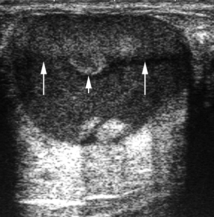
B
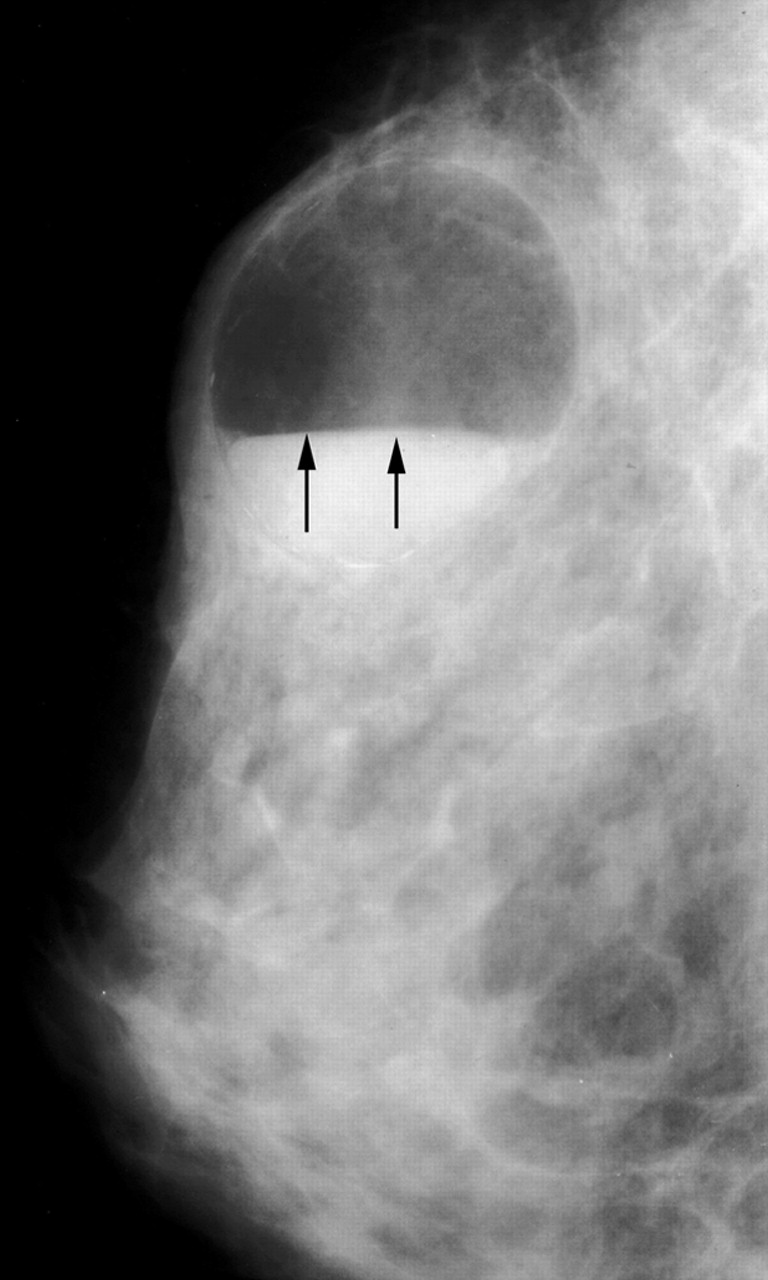
Cystic mass with fat-fluid level galactocele. (a) Mammogram reveals an oval circumscribed mass with the characteristic fat-fluid level (arrows). In this type of galactocele, the milk content is fresh and fluid, allowing the fat to rise and the heavier water content to remain in the lower portion of the cyst. (b) US image also demonstrates the fat-fluid level (long arrows), with typical high and low echogenicity. Note that the fatty component has risen and occupies the upper (nondependent) portion of the cyst, whereas the heavier water content remains in the lower (dependent) portion. Note also the clot of fatty milk (“cream”) (short arrow) floating in the nondependent portion of the cyst owing to its intermediate density.
http://radiographics.rsna.org/content/27/suppl_1/S101.figures-only
breast,duct ectasia
 Duct ectasia in the right breast in a 13-year-old girl. US scan depicts anechoic tubular structures.
http://www.google.com.eg/imgres?imgurl=http://radiographics.rsna.org/content/20/6/1613/F22.large.jpg&imgrefurl=http://radiographics.rsna.org/content/20/6/1613/F22.expansion&usg=__N3ecBH95OaAq2b_NcwMQZrDhtOw=&h=1456&w=1800&sz=321&hl=ar&start=1&sig2=txBavFP3CTgnpLBEf23IZw&zoom=1&itbs=1&tbnid=IgjqiXE3BF686M:&tbnh=121&tbnw=150&prev=/images%3Fq%3Dduct%2Bectasia,breast%26hl%3Dar%26sa%3DG%26gbv%3D2%26tbs%3Disch:1&ei=2mbuTMDcK9GB4Qam6anHCg
breast,duct ectasia
 This magnification left mediolateral galactogram demonstrates diffuse mild dilatation of the ducts. There were no persistent filling defects.
http://www.google.com.eg/imgres?imgurl=http://download.imaging.consult.com/ic/images/S1933033207712806/gr8-midi.jpg&imgrefurl=http://imaging.consult.com/image/topic/dx/Breast%3Ftitle%3DDuct%2520Ectasia%2520(Breast)%26image%3Dfig8%26locator%3D%26pii%3DS1933-0332(07)71280-6&usg=__90pbK4AcnglA5j71K6x70UxHq1g=&h=185&w=200&sz=4&hl=ar&start=13&sig2=0kdrB08PCwl4rO0AciPj9Q&zoom=1&itbs=1&tbnid=bAQUJoHIlrduXM:&tbnh=96&tbnw=104&prev=/images%3Fq%3Dduct%2Bectasia,breast%26hl%3Dar%26sa%3DG%26gbv%3D2%26tbs%3Disch:1&ei=AmbuTOzKB8SB4Qa5poWkCg
breast,intra-ductal papilloma
 Duplex Doppler images of the left subareolar region show that the intraluminal mass has arterial flow.
http://radiology.rsna.org/content/210/3/795.figures-only
breast,intra-ductal papilloma
 Transverse sonogram obtained with a 7.5-MHz linear transducer in the subareolar region. The curved arrow indicates the intraluminal solid mass. The straight arrows indicate the dilated duct.
http://radiology.rsna.org/content/210/3/795.figures-only
breast,intra-ductal papilloma
 Transverse sonograms obtained with a 7.5-MHz linear transducer in the subareolar region. The straight arrows show a dilated tubular structure. The curved arrow indicates an intraluminal solid mass. NIP refers to the location of the inverted nipple.
http://radiology.rsna.org/content/210/3/795.figures-only
Subscribe to:
Posts (Atom)




%26image%3Dfig5%26locator%3Dgr5%26pii%3DS1933-0332(07)71284-3&usg=__FJdizOtAQIWDi605CFKRgio-0wE=&h=200&w=174&sz=4&hl=ar&start=14&sig2=PnYic1maQ5WmAp0Ske2T9g&zoom=1&itbs=1&tbnid=M8eCBdRoQW4lxM:&tbnh=104&tbnw=90&prev=/images%3Fq%3Dbreast%2B,radial%2Bscar%26hl%3Dar%26sa%3DG%26gbv%3D2%26tbs%3Disch:1&ei=DAzwTMalNo_qOfilzPoJ)
%26image%3Dfig5%26locator%3Dgr5%26pii%3DS1933-0332(07)71275-2&usg=__GGlXaFZXLVESuRAP_3Wa4GxZm5A=&h=200&w=194&sz=7&hl=ar&start=1&sig2=9YhcxLxWCD8_KyJ_4waZog&zoom=1&itbs=1&tbnid=fIB9is-QR5OICM:&tbnh=104&tbnw=101&prev=/images%3Fq%3Dbreast%2Bhamartoma,ultrasound%26hl%3Dar%26sa%3DG%26gbv%3D2%26tbs%3Disch:1&ei=8ArwTNL-IsGEOtyjuYMK)


%26image%3Dfig3%26locator%3Dgr3%26pii%3DS1933-0332(07)71289-2&usg=__Xxezrq-JmuA9bY_ir4smb3EdGM4=&h=200&w=190&sz=4&hl=ar&start=1&sig2=NuP5-leUiMRhwNR-4ch6mg&zoom=1&itbs=1&tbnid=LIjuQlk-s5spbM:&tbnh=104&tbnw=99&prev=/images%3Fq%3Dlipoma,breast%26hl%3Dar%26sa%3DG%26gbv%3D2%26tbs%3Disch:1&ei=icbuTOPwL4iChQfMmYm5Cg)

%26image%3Dfig8%26locator%3D%26pii%3DS1933-0332(07)71280-6&usg=__90pbK4AcnglA5j71K6x70UxHq1g=&h=185&w=200&sz=4&hl=ar&start=13&sig2=0kdrB08PCwl4rO0AciPj9Q&zoom=1&itbs=1&tbnid=bAQUJoHIlrduXM:&tbnh=96&tbnw=104&prev=/images%3Fq%3Dduct%2Bectasia,breast%26hl%3Dar%26sa%3DG%26gbv%3D2%26tbs%3Disch:1&ei=AmbuTOzKB8SB4Qa5poWkCg)