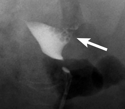
Hysterosalpingogram in 40-year-old woman shows medium-sized cesarean section scar defect arising from left lower uterine cavity wall (arrow).
http://www.ajronline.org/cgi/content/full/190/4/870/FIG1


 Hysterosalpingogram in 40-year-old woman shows medium-sized cesarean section scar defect arising from left lower uterine cavity wall (arrow).
 Tubal polyp. Spot radiograph shows a small filling defect (arrow) in the proximal left fallopian tube, a finding that typically represents a tubal polyp.
 a
a b
b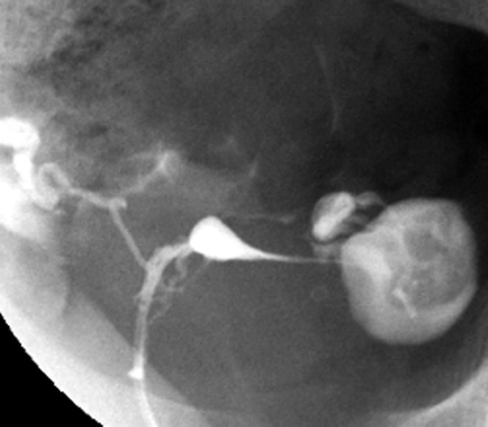 Peritubal adhesions. Spot radiograph demonstrates a round collection of contrast material adjacent to the left fallopian tube, a finding that suggests peritubal adhesions. Note the free contrast material spillage on the right side.
 a
a
 a
a b
b
 Cornual spasm. (a) On an HSG spot radiograph obtained during the early filling stage of the uterus, the right fallopian tube does not opacify beyond the cornual portion (arrow), whereas the left fallopian tube opacifies to the ampullary portion. Arrowheads indicate amorphous calcifications on the right side of the pelvis. These calcifications were also present on the scout image (not shown). (b) On a spot radiograph obtained after the instillation of additional contrast material, the right fallopian tube opacifies to the ampullary portion. Right-sided SIN and a left-sided hydrosalpinx are also noted. Amorphous calcifications (arrowheads) are again seen on the right side of the pelvis.
  SIN. Spot radiograph demonstrates SIN as small outpouchings or diverticula from the isthmic portion of the fallopian tubes. SIN can be either unilateral or (as in this case) bilateral.
 Cesarean section scar. Spot radiograph shows the uterine incision from a cesarean section (arrows) in the typical location (ie, oriented transverse in the lower uterine segment in the region of the isthmus). At HSG, a cesarean section scar can have a linear appearance (as in this case) or can occasionally manifest as a wedge-shaped outpouching or diverticulum.
 a
a b
b c
c d
d a
a b
b c
c
 Endometrial polyp. Sagittal sonohysterogram shows a large, well-defined mass in the fundus arising from the anterior aspect of the endometrium. Note the cystic area in the lower portion of the polyp.
 Uterine folds. HSG spot radiograph demonstrates uterine folds (arrows) as linear filling defects that parallel the longitudinal axis of the uterus. Uterine folds are normal findings that are occasionally seen at HSG.
 Septate and arcuate uterus. Spot radiograph demonstrates a depression of the uterine fundus, a finding that may represent a short septum or an arcuate deformity.
 Bicornuate uterus. Spot radiograph shows two markedly splayed uterine horns. The fallopian tubes are not visualized at this imaging stage.
 Hysterosalpingography (left image): The contrast material passes the tube from a single unicornate uterus (arrow) to the peritoneal cavity.