Best Bath Store
All Natural Blend of 14 Herbs Gentle Cleanse- $20 Off Colonix Now! Use Promo Code 20J
Amberen - Breakthrough Menopause Treatment
728x90
Custom Search
Showing posts with label MULTI-SYSTEM DISEASE. Show all posts

Thursday, October 8, 2009

Leukaemia


Leukaemia.
Leukaemia is a process involving the uncontrolled proliferation of the blood forming cells. The name of the particular leukaemia comes from the type of white cells that proliferate. Theories of its generation involve the idea of delayed maturation. That is if an ancestor cell in the bone marrow goes thorugh a few more cell divisions before forming the recognisable precursor adult cell then the number of those cells will increase. Research into the multiple factors in cell genetics is proceeding so fast that it is more appropriate to recommend the nearest search engine to enquiring minds.
The visible evidence of Leukamia usually follows an aggressive or a long standing abnormality. The radiologic expression of the disease is of an enlargement of the soft tissue mass of the bone marrow with loss of bone, particularly at the metaphyses where bone turnover is a little higher. The bone cortex amy also be eroded from below, giving a scalloped appearance. There may be other clues, such as splenic anlargement, visible on a radiograph of the pelvis. Lytic areas, chloromas, are rare, but are due to local masses of leukaemic cells.
The stem cell theory suggests that some cases of myelofibrosis might be included in this document, but the
condition is commoner in polycythaemia.


Figure 3:Clinical presentation:Adult male with anaemia.
The particular feature is the low density of pubis and sacrum with relative sparing of ilia and femora. Patchy bone loss can be seen in both ischia and the proximal metaphyses of both femora, coarsening the visible bone architecture. There is a fairly well-defined area of patchy bone loss in the upper part of each pubic bone centred around the mid-line. Some cortical scalloping of the cortex of pubic bones is present. The density of the ilia is probably normal, except adjacent to the sacro-iliac joint margins where some more extensive destruction is seen on the right side. The density of Sacrum and vertebrae is reduced by what appears to be a patchy destructive process.
The appearance is of a patchy lytic bone process that is predominatly mid-line. This might suggest a process that affects active bone marrow, which is often restricted in the adult to the axial skeleton and the medulla of adjacent parts of long bones.
D=lk inf


Wednesday, October 7, 2009

Lymphoma overview


Click on the image to enlarge


Lymphoma overveiw
Lymphoma is a type of cancer
involving cells of the immune system, called lymphocytes. Just as cancer represents many different diseases, lymphoma represents many different cancers of lymphocytes-about 35 different subtypes, in fact.
Lymphoma is a group of cancers that affect the cells that play a role in the immune system, and primarily represents cells involved in the lymphatic system
of the body.


The lymphatic system is part of the immune system. It consists of a network of vessels that carry a fluid called lymph, similar to the way that the network of blood vessels carry blood throughout the body. Lymph contains white blood cells called lymphocytes. Lymphocytes attack a variety of infectious agents as well as many cells in the precancerous
stages of development.
Lymph nodes are small collections of lymph tissue that occur throughout the body. The lymphatic system involves lymphatic channels that connect thousands of lymph nodes scattered throughout the body. Lymph flows through the lymph nodes, as well as through other lymphatic tissues including the spleen
, the tonsils, the bone marrow, and the thymus gland.
These lymph nodes filter the lymph, which may carry bacteria, viruses, or other microbes. The lymph nodes, or glands as they may be called, filter the lymph, which may on various occasions carry different microbial organisms. At infection sites, large numbers of these microbial organisms collect in the regional nodes and produce the swelling and tenderness typical of a localized infection. These enlarged and occasionally confluent collections of lymph nodes (so-called lymphadenopathy
) are often referred to as "swollen glands."
Lymphocytes recognize pathogens (infections and abnormal cells) and destroy them. There are 2 major subtypes of lymphocytes: B lymphocytes and T lymphocytes, also referred to as B cells and T cells.
B lymphocytes produce antibodies (proteins that circulate through the blood and lymph and attach to infectious organisms and abnormal cells). The combination attachment cell or antibody
microbial organism essentially alerts other cells of the immune system recognize and destroy these intruders, also known as pathogens.
T cells, when activated, can kill pathogens directly. T cells also play a part in the mechanisms of immune system control, to prevent the system from inappropriate overactivity or underactivity.
After fighting off an invader, some of the B and T lymphocytes "remember" the invader and are prepared to fight it off if it returns.
Cancer occurs when normal cells undergo a transformation whereby they grow and multiply uncontrollably. Lymphoma is a malignant
transformation of either lymphocytes B or T cells or their subtypes.
As the abnormal cells multiply, they may collect in 1 or more lymph nodes or in other lymph tissues such as the spleen.
As the cells continue to multiply, they form a mass often referred to as a tumor
.
Tumors often overwhelm surrounding tissues by invading their space, thereby depriving them of the necessary oxygen
and nutrients needed to survive and function normally.
Because of their uncontrolled growth, lymphomas can encroach on and/or invade neighboring tissues or distant organs.
In lymphoma, abnormal lymphocytes travel from one lymph node
to the next, and sometimes to remote organs, via the lymphatic system.
While lymphomas are often confined to lymph nodes and other lymphatic tissue
, they can spread to other types of tissue almost anywhere in the body. Lymphoma development outside of lymphatic tissue is called extranodal disease.
Lymphomas fall into 1 of 2 major categories. Hodgkin lymphoma (HL, previously called Hodgkin's disease) and all other lymphomas (non-Hodgkin lymphomas or NHLs).
These 2 types occur in the same places, may be associated with the same symptoms, and often have similar gross physical characteristics. However, they are readily distinguishable via microscopic
examination.
Hodgkin disease develops from a specific abnormal B lymphocyte lineage. NHL may derive from either abnormal B or T cells and are distinguished by unique genetic markers.
There are 5 subtypes of Hodgkin disease and about 30 subtypes of non-Hodgkin lymphoma.
Because there are so many different subtypes of lymphoma, the classification of lymphomas is complicated and includes both the microscopic appearance and well-defined genetic and molecular rearrangements.
Many of the NHL subtypes look similar, but they are functionally quite different and respond to different therapies with different probabilities of cure. HL subtypes are microscopically distinct, and typing is based upon the microscopic differences as well as extent of disease.
Lymphoma is the most common type of blood cancer in the United States. It is the sixth most common cancer in adults and the third most common in children. Non-Hodgkin lymphoma is far more common than Hodgkin disease.
In the United States, about 54,000 new cases of NHL and 7000 new cases of HL were diagnosed in 2004, and the overall incidence is increasing.
About 24,000 people die of NHL and 1400 of HL each year, with the survival rate of all but the most advanced cases of HL greater than that of other lymphomas.
Lymphoma can occur at any age, including childhood. Hodgkin disease is most common in 2 age groups: young adults aged 16-34 years and in older people aged 55 years and older. Non-Hodgkin lymphoma is more likely to occur in older people.
PET/CT FINDINGS
Marked FDG uptake throughout the mediastinum and in the right axilla/supraclavicular area corresponding to bulky adenopathy on the CT portion of the exam compatible with malignancy.

Please see the lower image .

TREATMENT / FOLLOW UP
Chemotherapy (CHOP). Follow-up PET/CT ordered following 1 cycle.
FOLLOW UP PET/CT FINDINGS
Complete resolution of abnormal FDG activity compatible with a good response to therapy. Focal apparent FDG activity in the left supraclavicular area was not present on the uncorrected images compatible with an attenuation correction artifact. Bulky adenopathy is still present, but no increased FDG activity is present. Please see the upper image .
DISCUSSION
This case demonstrates the power of PET/CT to assess response to therapy soon after initiation. The strength of the modality is in the ability to assess an early response to therapy by assessing the metabolic changes. As shown in the second set of images, there is still considerable soft tissue abnormality present, but no increased FDG activity. Evidence suggests that for non-Hodgkin’s lymphoma, patients are to be categorized as responders (better overall survival) only if there is minimal or no residual FDG activity on follow up exams after therapy initiation. The metabolic changes can be assessed after one cycle of chemotherapy, whereas the soft tissue component will take much longer to regress and may remain indefinitely.

Wegeners granulomatosis

Click on image to enlarge


Wegener's Granulomatosis


General Considerations
Hallmarks
Small to medium sized systemic vasculitis
Granulomatous inflammation
Necrosis
Unknown etiology
Incidence of 1 case per 30,000
Affects predominantly whites (91%)
M>F
Mean age at diagnosis = 45

Sites of Involvement:
Lung (>90%)
Renal (75%)
Rapidly progressing glomerulonephritis, chronic renal failure
Trachea (15-60%)
Subglottic Stenosis
Other
Paranasal sinuses, nose (saddle nose deformity)
Ears, eyes, oral cavity
Skin, joints, nervous system
Rarely
Heart, GI tract and brain
Can potentially involve any organ in the body
Diagnosis:
American College of Rheumatology Classification
(2 out of 4 criteria is 88% sensitive and 92% specific for the diagnosis)
Nasal or oral inflammation
Abnormal chest X-ray
Urinary sediment
Biopsy
Imaging Findings
Conventional Radiography
Pulmonary nodules
Most common finding (40-70%)
Typically multiple and bilateral with a tendency to cavitate (50%)
Cavitary lesions may lead to atelectasis or pneumothorax
Thick or thin walled, well or ill circumscribed
Tendency to wax and wane
Size varies (1.5-10cm)
Air space consolidation
Waxing and waning infiltrates
May be mistaken for pneumonia
Pulmonary hemorrhage or edema
Hilar lypmhadenopathy
Normal in 20%
CT
Useful in further defining extent of disease seen in plain film, and revealing lesions not seen on plain film including:
Interstitial abnormalities
Tracheobronchial Abnormalities
Findings suggestive of vasculitis
Differential Diagnosis
For cavitary lung lesions
Infarction
Septic pulmonary emboli
Carcinoma
Squamous cell carcinoma
Infection
TB, Fungal, Bacterial
Rheumatoid nodules
Prognosis and Treatment
Tracheostomy may be required for tracheal strictures
Independent Risk factors of mortality
Older age
Absence of ear, nose and throat involvement
Renal or Cardiac involvement


Langerhans histiocytosis

Click on image to enlarge

Langerhans Histiocytosis

Also known as eosinophilic granuloma(tosis)
Proliferative disorder of the Langerhans cells
Normally found in the skin (and a few other organs) and serve as antigen-presenting cells
Rare diseases, affecting neonates up to adults
2:1 male to female predominance
Prognosis
Mortality and morbidity are associated with the clinical presentation and age of onset of the disease
Worst prognosis for neonates presenting with the disseminated form
Three clinical forms
Acute disseminated Langerhans cell histiocytosis (aka Letterer-Siwe disease)
Occurs most frequently in infants 2 years of age or younger (and occasionally adults)
Presents with multi system organ involvement
Cutaneous lesions resembling seborrheic dermatitis involve the scalp, face, trunk and buttocks as the dominant clinical feature (nearly 80% of patient will have this)
Infiltration of bone marrow and other organs lead to concurrent hepatosplenomegaly, lypmhadenopathy, pulmonary lesions, anemia, thrombocytopenia, recurrent infections (otitis media)
Eventually, there are destructive osteolytic bone lesions
If untreated, this disease is rapidly fatal
With chemotherapy, 5 year survival rate is approximately 50 percent
Univocal Langerhans cell histiocytosis (aka Eosinophilic granuloma or granulomatosis)
Usually only affects the skeletal system of young adults
Typically presents as an osteolytic lesion involving the
Calvaria
Vertebra
Rib
Mandible
Femur
Ilium
Scapula
Bony lesions are usually asymptomatic
In some cases, can cause pain and even pathologic fractures
Pulmonary lesions may be the only presenting symptom and organ involved, especially in adults
Skeletal lesion is usually indolent in nature
Can heal spontaneously or be cured by local excision or irradiation
Pulmonary lesions are typically followed and treated with supportive care
Multifocal Langerhans cell histiocytosis (aka Hand-Schuller-Christian disease)
Triad
Diabetes insipidus
Exophtalmos
Holes in the bone, usually the head (calvarium)
Commonly affects children
Can lead to
Lypmhadenopathy
Hepatomegaly
Splenomegally
Diabetes insipidus is secondary to infiltration of the posterior pituitary stalk by the Langerhans cell
About a third of these patients will also display cutaneous lesions
Some will experience spontaneous regression while others can be treated with chemotherapy


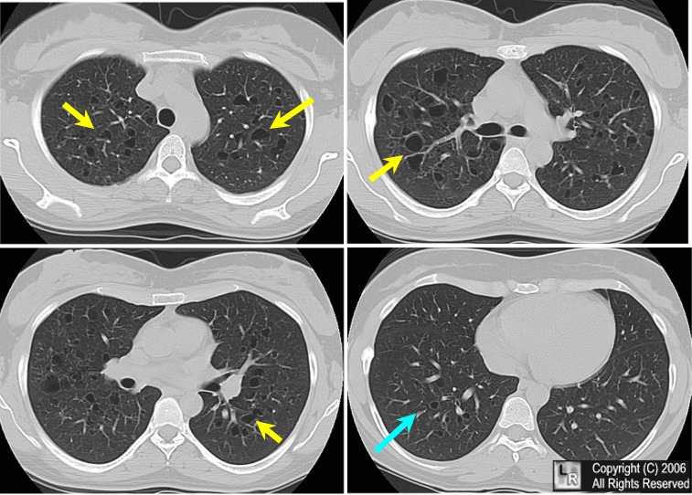
Langerhans histiocytosis (Eosinophilic granuloma of lung). Four selected non-enhanced axial CT scans of the chest show multiple small, irregularly-shaped, cysts of varying sizes with thin walls scattered throughout the lungs (yellow arrows) but predominantly seen in the upper lung fields while sparing the costophrenic angles and lung bases (blue arrow).

Quick Facts

Letterer-Siwe Disease
10% of histiocytosis X
Acute disseminated, fulminant form
Age at onset
Several weeks after birth to 2 years
Pathology
May be confused with leukemia
Symptoms
Hemorrhage, purpura
Severe anemia
Fever
Hepatosplenomegaly and lypmhadenopathy
Bone involvement in 50%
Widespread lytic lesions
Prognosis: 70% mortality rate
Hand-Schuller-Christian
15-40% of Histiocytosis X
Triad of:
Exophthalmos (33%)
Diabetes insipidus (30-50%)
Lytic skull lesions
Pathology
May simulate Ewing's sarcoma
Age at onset
5-10 years
Target organs
Bone
Lytic skull lesions with overlying soft tissue nodules
Large geographic skull lesions
"Floating teeth" with mandibular involvement
Soft tissue
Hepatosplenomegaly is rare
Lypmhadenopathy which may be massive
Lung
Cyst and bleb formation with spontaneous PTX
Ill-defined diffuse nodular disease often leading to fibrosis and honeycombing
Prognosis: spontaneous remissions and exacerbations
Eosinophilic granuloma
60-80% of Histiocytosis X
Usually confined to bone
Age at onset
5-10 years highest frequency
Male predominance 3:2
Location
Calvarium>mandible>spine>ribs>long bones
Most are monostotic (50-75%)
Target organs
Skull (50%)
Diploic space of parietal bone most often
Round or ovoid punched out lesions with beveled edge
Sclerotic margin during healing phase
Beveled edge=hole-within-a-hole
Button sequestrum- bony sequestrum within lytic lesion
Axial skeleton (25%)
"Vertebra plana"-"coin-on-edge"(Calve disease)=collapse of vertebral body, mostly thoracic
Most common cause of vertebra plana in children
Proximal long bones (15%)
Expansile, lytic lesions, mostly diaphyseal
Soft tissue mass
Laminated periosteal reaction
Lung (20%)
Age peak between 20-40 years
Multiple small nodules
Predilection for apices
Prototype for honeycomb lung
Recurrent pneumothoraces (25%)
Rib lesions with fractures common
Nuclear Medicine
Negative bone scans in 35%
Bone lesions usually not Ga-67 avid
Ga-67 may be helpful in detecting non-osseous lesions
Prognosis: excellent