


The major categories of the FIGO classification are as follows:
Stage 0 – Carcinoma in situ
Stage I – Invasive carcinoma that is strictly confined to the cervix
Stage II – Locoregional spread of the cancer beyond the uterus but not to the pelvic sidewall or the lower third of the vagina
Stage III – Cancerous spread to the pelvic sidewall or the lower third of the vagina, and/or hydronephrosis or a nonfunctioning kidney that is incident to invasion of the ureter
Stage IV – Cancerous spread beyond the true pelvis or into the mucosa of the bladder or rectum
The FIGO stages are further categorized as follows:
Stage Ia cervical carcinoma – Preclinical invasive carcinoma that can be diagnosed only by means of microscopy
Stage Ib cervical carcinoma – A clinically visible lesion that is confined to the cervix uteri
Stage Ib1 – The primary tumor is not greater than 4.0 cm in diameter.
Stage Ib2 – The primary tumor is greater than 4.0 cm in diameter.
Stage IIa cervical carcinoma – Spread into the upper two thirds of the vagina without parametrial invasion
Stage IIb cervical carcinoma – Extension into the parametrium but not into the pelvic sidewall
Stage IIIa cervical carcinoma – Extension into lower one third of the vagina, without spread to the pelvic sidewall
Stage IIIb cervical carcinoma – Extension into the pelvic sidewall and/or causes a nonfunctioning kidney or hydronephrosis due to invasion of the ureter
Stage IVa cervical carcinoma – Extension of the tumor into the mucosa of the bladder or rectum
Stage IVb cervical carcinoma – Spread of the tumor beyond the true pelvis and/or by metastasis into distant organs
The strict FIGO clinical staging guidelines do not include the status of the lymph nodes, although the presence of metastatic adenopathy is an important factor in treatment planning and in the prognosis.
Extended clinical staging with cross-sectional imaging (CT scanning and/or MRI) includes the status of the lymph nodes in the assessment of the extent of the disease. The detection of enlarged pelvic lymph nodes is considered equivalent to pelvic sidewall tumor extension (stage III), and the detection of enlarged lymph nodes in the para-aortic, paracaval, or inguinal regions is considered extrapelvic tumor spread (stage IV).
The major limitations of the FIGO clinical staging system are encountered in the estimation of the size of the primary tumor, particularly when the tumor is endocervical. The size of the tumor is significant because, in each stage, the incidence of lymph node metastases increases and the prognosis deteriorates with increased volume of the primary tumor.
Other limitations occur in the evaluation of tumor extension into the parametrium and pelvic sidewalls and in the detection of metastatic lymphadenopathy or distant metastasis.Extended clinical staging with cross-sectional imaging (CT scanning and/or MRI) and surgicopathologic staging, including pelvic and abdominal retroperitoneal lymphadenectomy, provide additional diagnostic value.
Each has been proven to be superior to the conventional FIGO clinical staging system in determining the full extent of the tumor spread. However, once the clinical stage is assigned on the basis of the clinical pretreatment workup results (in compliance with the FIGO guidelines), the stage should not be altered as a result of subsequent findings. Instead, any additional information that is revealed by cross-sectional imaging or surgery is primarily used for planning treatment regimens, and they should not be used to revise the assigned clinical stage.
http://emedicine.medscape.com/article/402329-media
Showing posts with label cervical carcinoma. Show all posts
Showing posts with label cervical carcinoma. Show all posts
Wednesday, June 30, 2010

Human papillomavirus (HPV) is now recognized as the most important causative agent in cervical carcinogenesis at the molecular level, although HPV may not induce many of the identified molecular alterations.11,12 As many as 5% of cervical cancers may not be associated with HPV.13,14 First intercourse at an early age, sexual promiscuity, high parity, race, and low socioeconomic status are presently thought to increase the risk for cervical cancer because these factors are linked to sexual behavior that increases the likelihood of exposure to HPV and/or because they are cofactors that modify the risk in women who are infected with HPV. Tobacco smoking is also a significant independent risk factor.
http:// emedicine.medscape.com/article/402329-media
http:// emedicine.medscape.com/article/402329-media
spread of cervical carcinoma
1-The main pathways for the spread of invasive cervical cancer consist of the following: (1) microscopic spread into the vaginal mucosa beyond a visible or palpable tumor; (2) extension into the endometrium or myometrium of the corpus; (3) direct extension into the parametrium and, in advanced stages, into the adjacent structures; and (4) spread into the regional pelvic lymph nodes and, potentially, into the retroperitoneal, inguinal, or thoracic lymph nodes.
2-The extrauterine spread of cervical cancer occurs primarily by means of direct extension and lymphatic invasion that initially affects the contiguous tissues in the region of the laterally positioned cardinal ligament. Later, the tumor may involve the anterior or posterior parametrium, which are separated from the cervix by the pubovesicocervical fascia. The lateral paracervical and parametrial regions are more vulnerable to tumor invasion than the anterior and posterior parametrium because of the lack of a protective fascial covering at the lateral regions and because of the natural lymphatic drainage through the lateral paracervical tissues into the cardinal ligaments.
3-The local spread of cervical cancer may progress through the parametrium to involve the ureters and, eventually, the pelvic sidewalls. In some patients, the sciatic plexus is involved. Hydronephrosis, pyelonephritis, and renal failure are common complications of progressive disease. Involvement of the urinary bladder and rectum can occur in advanced cases because of direct tumor extension or subsequent to invasion of the vesicouterine or uterosacral ligaments, respectively. A vesicovaginal fistula or rectovaginal fistula may or may not develop. A tumor that extends through the posterior aspect of the cervix or corpus infrequently leads to intraperitoneal spread. Adnexal metastases are uncommon intheearlystages of the disease.
4-Lymphatic tumor spread usually occurs in a fairly orderly pattern or sequence that first involves the regional paracervical and parametrial lymph nodes and then the internal and external iliac lymph nodes. This may then be followed by spread to the common iliac nodes, the para-aortic nodes, and eventually the supraclavicular nodes via the thoracic duct. Metastasis to the para-aortic lymph nodes without involvement of pelvic lymph nodes is unusual.
5-Hematogenous tumor spread may be a result of a lymphatic venous anastomosis or direct venous invasion. The most common sites of hematogenous metastases are the lungs, bones, and liver.
http://emedicine.medscape.com/article/402329-media
1-The main pathways for the spread of invasive cervical cancer consist of the following: (1) microscopic spread into the vaginal mucosa beyond a visible or palpable tumor; (2) extension into the endometrium or myometrium of the corpus; (3) direct extension into the parametrium and, in advanced stages, into the adjacent structures; and (4) spread into the regional pelvic lymph nodes and, potentially, into the retroperitoneal, inguinal, or thoracic lymph nodes.
2-The extrauterine spread of cervical cancer occurs primarily by means of direct extension and lymphatic invasion that initially affects the contiguous tissues in the region of the laterally positioned cardinal ligament. Later, the tumor may involve the anterior or posterior parametrium, which are separated from the cervix by the pubovesicocervical fascia. The lateral paracervical and parametrial regions are more vulnerable to tumor invasion than the anterior and posterior parametrium because of the lack of a protective fascial covering at the lateral regions and because of the natural lymphatic drainage through the lateral paracervical tissues into the cardinal ligaments.
3-The local spread of cervical cancer may progress through the parametrium to involve the ureters and, eventually, the pelvic sidewalls. In some patients, the sciatic plexus is involved. Hydronephrosis, pyelonephritis, and renal failure are common complications of progressive disease. Involvement of the urinary bladder and rectum can occur in advanced cases because of direct tumor extension or subsequent to invasion of the vesicouterine or uterosacral ligaments, respectively. A vesicovaginal fistula or rectovaginal fistula may or may not develop. A tumor that extends through the posterior aspect of the cervix or corpus infrequently leads to intraperitoneal spread. Adnexal metastases are uncommon intheearlystages of the disease.
4-Lymphatic tumor spread usually occurs in a fairly orderly pattern or sequence that first involves the regional paracervical and parametrial lymph nodes and then the internal and external iliac lymph nodes. This may then be followed by spread to the common iliac nodes, the para-aortic nodes, and eventually the supraclavicular nodes via the thoracic duct. Metastasis to the para-aortic lymph nodes without involvement of pelvic lymph nodes is unusual.
5-Hematogenous tumor spread may be a result of a lymphatic venous anastomosis or direct venous invasion. The most common sites of hematogenous metastases are the lungs, bones, and liver.
http://emedicine.medscape.com/article/402329-media

 Computed tomography (CT) scan of clinical stage IIb cervical carcinoma (same patient in Images 4-5 in Multimedia). The parametrial invasion is depicted with CT scanning as loss of definition of the cervical contours, accompanied by increased attenuation and prominent soft-tissue stranding in the parametrial fat. Parametritis can result in similar findings. The cervix shows ill-defined hypoattenuation, but the tumor is not clearly delineated. In addition, a subserosal leiomyoma protrudes from the left side of the uterus.
 Computed tomography scan of a large, lobulated mass that is replacing the cervix and showing nonuniform hypoattenuation. The air and fluid in the center of the mass are consistent with tumor necrosis and a complicating infection (the patient had purulent discharge). The central hypoattenuation in the uterine corpus is suggestive of minimal fluid in the cavity.
http:// emedicine.medscape.com/article/402329-media

 Computed tomography scan through the upper uterus (same patient in Images 1-2 in Multimedia). This image shows fluid that markedly distends the endometrial cavity secondary to obstruction of the endocervical canal by cervical cancer. A small submucosal leiomyoma projects into the right anterior aspect of the endometrial cavity and has minute calcifications.
http://%20emedicine.medscape.com/article/402329-media

 Computed tomography (CT) scan of parametrial and rectal invasion by cervical carcinoma (same patient in Images 10-11 in Multimedia). There is loss of definition of the cervical contours, accompanied by a masslike soft tissue that replaces the parametrial fat on the right and that extends into the anterior and right-sided rectal walls
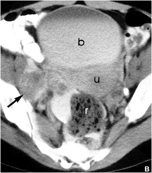
 This computed tomography scan demonstrates a markedly enlarged lymph node at the left pelvic sidewall, a finding that is consistent with pelvic lymph node metastasis, which is indicative of stage IIIb disease. The cystic consistency is not unusual for metastatic cervical carcinoma. The primary tumor is well depicted as a hypoattenuating, circumscribed mass. A cyst is present in the anteriorly located left ovary.
http://emedicine.medscape.com/article/402329-media
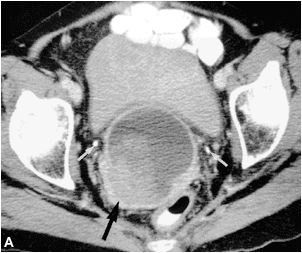
 This computed tomography scan demonstrates a cervical tumor directly extending into the posterior wall of the bladder and into the left pelvic sidewall. Extension into the pelvic sidewall is a feature of stage IIIb disease, whereas involvement of the bladder wall is a feature of stage IVa disease.
http:// emedicine.medscape.com/article/402329-media


 Computed tomography (CT) scan of stage IVb cervical carcinoma (same patient in Images 14-17 in Multimedia). There is the presence of borderline enlarged para-aortic and pelvic lymph nodes, presumably secondary to metastasis; left hydronephrosis and hydroureter to the level of the large cervical tumor; and direct intraluminal extension of the tumor into the bladder. The borderline enlarged left para-aortic lymph node is consistent with stage IVb cervical carcinoma.
Sagittal T2-weighted magnetic resonance image (MRI) of a large cervical tumor extending through the entire cervical stroma and extensively involving the uterine corpus, vagina, bladder wall, and posterior urethral region . The invasion of the bladder wall is a feature of stage IVa disease. Courtesy of Kaori Togashi, MD, Hitachi Medical Corporation, Chair of Department of Diagnostic and Interventional Imageology, Kyoto University, Japan.
 Axial T2-weighted magnetic resonance image (MRI) of a large cervical tumor with full-thickness stromal invasion causing complete loss of the hypointense stromal stripe or ring (same patient in Images 22-23 in Multimedia). Also depicted is invasion of the parametrium and the posterior bladder wall; this finding is indicative of stage IVa disease. Courtesy of Kaori Togashi, MD, Hitachi Medical Corporation, Chair of Department of Diagnostic and Interventional Imageology, Kyoto University, Japan.
http:// emedicine.medscape.com/article/402329-media
 This sagittal transabdominal sonogram shows a circumscribed hypoechoic tumor in the posterior aspect of the cervix
http://emedicine.medscape.com/article/402329-media
 This transverse transvaginal sonogram shows a circumscribed hypoechoic tumor in the left posterior aspect of the cervix
http://emedicine.medscape.com/article/402329-media


Friday, April 16, 2010


Thursday, April 15, 2010


Subscribe to:
Posts (Atom)




