


ESSENTIALS
-Many patients with inflammatory or infectious types of colitis present with abdominal pain, and multidetector CT is often used as the initial diagnostic test.
-CT can accurately depict the bowel wall and help determine the presence and extent of intramural disease, its possible extraluminal extension, and involvement of distant sites.
-Many imaging features or patterns are helpful in distinguishing the various types of colitis, including degree of wall thickening, extent and location of disease, extraluminal manifestations, and types of complication.
-While imaging features can help narrow the differential diagnosis or suggest a specific diagnosis, the final diagnosis is often based on clinical and laboratory data and colonoscopic and biopsy findings.
radiology.rsna.org/content/240/3/623.full
Showing posts with label intestinal lesions. Show all posts
Showing posts with label intestinal lesions. Show all posts
Saturday, June 12, 2010

 Transverse CT image in a 34-year-old man with acute right lower quadrant pain caused by appendicitis with perforation. Appendiceal lumen (short black arrow) is enlarged and shows wall enhancement. Mesenteric stranding (white arrow), free intraperitoneal air (arrowhead), and thickened terminal ileum (long black arrow) due to contiguous inflammation are seen.
radiology.rsna.org/content/240/3/623.full
 Transverse CT image in a 21-year-old man with right lower quadrant pain due to appendicitis shows an appendicolith (arrowhead) separated from cecal lumen by cecal wall thickening (cecal bar sign) (black arrow). Dilatation of inflamed appendix with air and debris (white arrow) is demonstrated.
radiology.rsna.org/content/240/3/623.full

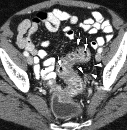
Transverse CT image in a 65-year-old woman with early diverticulitis who had left lower quadrant pain and leukocytosis reveals wall thickening and multiple small diverticula (arrows) in a long segment of the sigmoid colon. Fascial thickening (arrowheads) along left pelvic side wall indicates mild diverticulitis.
 a
a b
bIschemic colitis in an 81-year-old woman with myocardial infarction. (a) Anteroposterior CT scout view shows air (arrows) in wall of right colon and small- and large-bowel dilatation. (b) Transverse CT image demonstrates air (arrowheads) in wall of right colon, with lack of wall enhancement and pericolonic stranding indicative of infarction.
 Transverse CT image in a 22-year-old woman with ischemic colitis after blunt abdominal trauma to right flank demonstrates marked thickening of hepatic flexure and right colon, with abrupt transition (arrows) between abnormal and normal wall in the transverse colon.
radiology.rsna.org/content/240/3/623.full
 Transverse CT image in a 71-year-old man with ischemic colitis due to arrhythmia shows ascites and marked thickening of sigmoid colon associated with multiple large nodular defects, which are the CT analog of thumbprinting (arrows) on radiographs. Note that rectal wall (arrowheads) is normal.
radiology.rsna.org/content/240/3/623.full
 Transverse CT image in a 45-year-old male transplantation patient with neutropenic colitis shows marked wall thickening in ascending colon (arrow) associated with pericolonic stranding and ascites (arrowheads).
radiology.rsna.org/content/240/3/623.full
 Transverse CT image in a 9-year-old girl with myelogenous leukemia and typhlitis demonstrates marked wall thickening in cecum (arrow) and terminal ileum (arrowhead).
radiology.rsna.org/content/240/3/623.full

 a
a b
bTransverse CT images in a 31-year-old man with amebiasis who had a history of diarrhea and recent travel to North Africa. (a) Cecal wall is thickened (arrow), but terminal ileum (arrowheads) is not involved. (b) Hepatic flexure (arrows) also demonstrates marked thickening, but descending colon (arrowhead) appears normal.
 Transverse CT image in a 60-year-old man with fistula formation due to tuberculous colitis. Descending colon appears slightly thickened, and a fistulous tract (white arrows) is shown to extend from the colon to an abscess (black arrow) in the left psoas muscle and through the left paraspinal muscles into the subcutaneous tissue of the back, where another abscess (arrowheads) has formed.
radiology.rsna.org/content/240/3/623.full
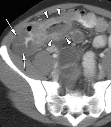 Transverse CT image in a 33-year-old man with tuberculous ileocolitis who had recently emigrated from Southeast Asia. The cecal wall (arrows) and terminal ileum (arrowheads) are markedly thickened.
radiology.rsna.org/content/240/3/623.full

 CT images in a 37-year-old woman with Crohn disease (ileocolitis). (a) Transverse scan obtained following enteroclysis demonstrates ileoileal (arrow) and ileosigmoidal fistulae (arrowhead). The involved ileal and colonic wall is asymmetrically thickened. (b) Coronal reformation shows ileoileal fistula (arrow) quite well. Contrast enhancement in vagina (arrowheads) is due to an ileovaginal fistula. (Images courtesy of John Lappas, MD, University of Indiana.)
  Transverse CT image in a 35-year-old patient with ulcerative colitis and toxic megacolon shows markedly distended transverse colon with shaggy mucosa (arrows).
radiology.rsna.org/content/240/3/623.full
 Transverse CT image in a 24-year-old woman with Crohn ileocolitis shows thickening of terminal ileum and cecum (white arrows) with fibrofatty proliferation (arrowheads) in right lower quadrant. One enlarged lymph node (black arrow) is also depicted.
radiology.rsna.org/content/240/3/623.full
 Transverse CT image in a 32-year-old woman with ulcerative colitis and bloody diarrhea demonstrates the double halo, or target, sign with inner (mucosa, arrow) and outer (muscularis propria, arrowhead) rings of high attenuation separated by a ring of low attenuation, which represents submucosa with edema. Little if any pericolonic stranding is seen.
http://www.google.com.eg/imgres?imgurl=http://radiology.rsna.org/content/240/3/623/F16.small.gif&imgrefurl=http://radiology.rsna.org/content/240/3/623.full&usg=__TEFfqDAl8UDupdZnziMD_QJcGvU=&h=187&w=200&sz=34&hl=en&start=25&itbs=1&tbnid=0LErWAgkUG2ioM:&tbnh=97&tbnw=104&prev=/images%3Fq%3Dleukaemia%2Bradiology%26start%3D20%26hl%3Den%26sa%3DN%26gbv%3D2%26ndsp%3D20%26tbs%3Disch:1
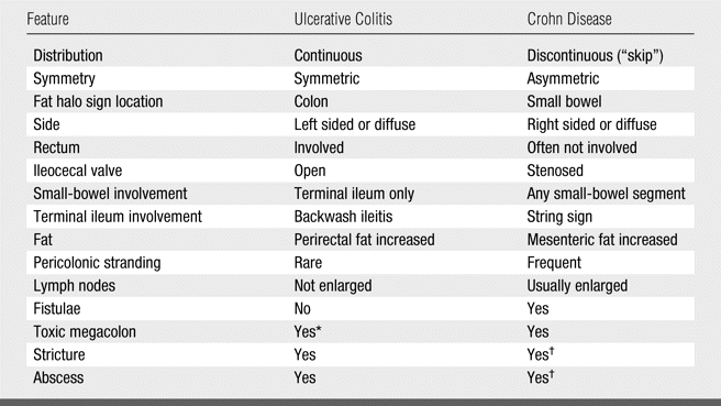 Distinction between Ulcerative Colitis and Crohn Disease
http://www.google.com.eg/imgres?imgurl=http://radiology.rsna.org/content/240/3/623/F16.small.gif&imgrefurl=http://radiology.rsna.org/content/240/3/623.full&usg=__TEFfqDAl8UDupdZnziMD_QJcGvU=&h=187&w=200&sz=34&hl=en&start=25&itbs=1&tbnid=0LErWAgkUG2ioM:&tbnh=97&tbnw=104&prev=/images%3Fq%3Dleukaemia%2Bradiology%26start%3D20%26hl%3Den%26sa%3DN%26gbv%3D2%26ndsp%3D20%26tbs%3Disch:1
 Transverse CT image in a 45-year-old man with chronic ulcerative colitis. Thickened rectal wall demonstrates the fat halo sign (arrowhead). Perirectal fat (arrows) is increased.
http://www.google.com.eg/imgres?imgurl=http://radiology.rsna.org/content/240/3/623/F16.small.gif&imgrefurl=http://radiology.rsna.org/content/240/3/623.full&usg=__TEFfqDAl8UDupdZnziMD_QJcGvU=&h=187&w=200&sz=34&hl=en&start=25&itbs=1&tbnid=0LErWAgkUG2ioM:&tbnh=97&tbnw=104&prev=/images%3Fq%3Dleukaemia%2Bradiology%26start%3D20%26hl%3Den%26sa%3DN%26gbv%3D2%26ndsp%3D20%26tbs%3Disch:1
Subscribe to:
Posts (Atom)




