


 Neck CT scan demonstrating large thyroid lymphoma
Showing posts with label lymphoma. Show all posts
Showing posts with label lymphoma. Show all posts
Sunday, June 13, 2010

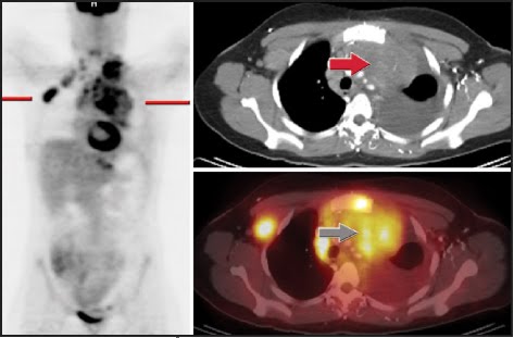 Marked FDG uptake throughout the mediastinum and in the right axilla/supraclavicular area corresponding to bulky adenopathy on the CT portion of the exam compatible with malignancy.
 Chemotherapy (CHOP). Follow-up PET/CT ordered following 1 cycle.
Complete resolution of abnormal FDG activity compatible with a good response to therapy. Focal apparent FDG activity in the left supraclavicular area was not present on the uncorrected images compatible with an attenuation correction artifact. Bulky adenopathy is still present, but no increased FDG activity is present.
Discussion:
This case demonstrates the power of PET/CT to assess response to therapy soon after initiation. The strength of the modality is in the ability to assess an early response to therapy by assessing the metabolic changes. As shown in the second set of images, there is still considerable soft tissue abnormality present, but no increased FDG activity. Evidence suggests that for non-Hodgkin’s lymphoma, patients are to be categorized as responders (better overall survival) only if there is minimal or no residual FDG activity on follow up exams after therapy initiation. The metabolic changes can be assessed after one cycle of chemotherapy, whereas the soft tissue component will take much longer to regress and may remain indefinitely.

 CT non contrast
 ADC MAP
 DWI
DWI Axial flair
 MR perfusion CBV map
findings:Multiple enhancing mass lesions with increased choline and restricted diffusion and minimally decreased perfusion.
D.D.:Metastases, abscesses, lymphoma, demyelination
Discusion:Lymphoma with high cellularity may show restricted diffusion and iso or slightly decreased perfusion.
 CT showed lymphadenopathy above and below the diaphragm and a lesion in L-3 vertebra consistent with a compressed or pathological fracture. The final diagnosis of lymphoma was made after biopsy of enlarged right inguinal lymph nodes and by bone marrow biopsy. There are areas of increased pathological activity in the left supra and infraclavicular region, the upper and mid-mediastinum, the right pulmonary hilus, D-8, D-12, L-2, L-3, L-4 vertebrae and the left inguinal region.
http://www.blogger.com/www.gehealthcare.com/.../products/lymphoma.html
 Contrast-enhanced helical CT scan demonstrates a markedly enlarged, conglomerate nodal mass with homogeneous attenuation enveloping the retroperitoneum. Lymphoma was the most likely diagnosis, but biopsy revealed adenocarcinoma.
radiographics.rsna.org/.../1/197/F38.expansion
orbital lymphoma

 This is 68 year old man with bilateral propotosis and lymphadenopathy. CT images are characterstic of orbital lymphoma.
 PET/CT for staging of Hodgkin's lymphoma. CT showed involvement only in right neck. PET/CT (A: coronal views; B: transverse views; MIP = maximum-intensity projection) showed that normal-size (9-mm) upper mediastinal lymph node was clearly metabolically active, changing stage from I to II. This finding is relevant if consolidative radiation after chemotherapy is planned. Incidental normal scalene muscle uptake was noted on coronal PET.
http://jnm.snmjournals.org/cgi/content-nw/full/48/1_suppl/19S/FIG1
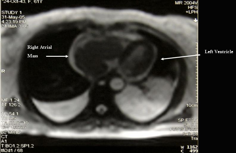 Primary right atrial lymphoma. Magnetic Resonance Imaging # Description : Magnetic Resonance Imaging of primary right atrial lymphoma
 Primary right atrial lymphoma. CT scan of jugular veins # Description : Contrast CT scan showing extension of the primary right atrial lymphoma through the superior vena cava up to the right jugular vein that is occluded while the contra-lateral jugular vein is opened.
 Figure 1. 55-year-old man with recurrent mantle cell lymphoma. CT shows symmetric circumferential thickening of the distal trachea (arrows).
 Figure 2. 55-year-old man with recurrent mantle cell lymphoma. Axial PET/CT shows intense activity in the suspected area around the distal trachea.
Figure 3. 55-year-old man with recurrent mantle cell lymphoma. Full body coronal reformatted PET scan shows abnormal intense tracheal uptake and other areas of expected uptake.


Subscribe to:
Posts (Atom)




70542-0&usg=__uTZTDQZoEVoHH4U-pWmSd-gqSmo=&h=194&w=200&sz=6&hl=en&start=16&itbs=1&tbnid=krD-8l90xPz2yM:&tbnh=101&tbnw=104&prev=/images%3Fq%3Dlymphoma,ct%26hl%3Den%26sa%3DG%26gbv%3D2%26tbs%3Disch:1)


