Best Bath Store
All Natural Blend of 14 Herbs Gentle Cleanse- $20 Off Colonix Now! Use Promo Code 20J
Amberen - Breakthrough Menopause Treatment
728x90
Custom Search
Showing posts with label chest diseases. Show all posts

Thursday, August 5, 2010

Kartagener's Syndrome with Situs Inversus on CXR and CT Scan

Findings
-The CXR demonstrates situs inversus with dextrocardia, right sided aorta, right gastric air bubble, left sided liver, and bronchiectasis. The CT confirms the dextrocardia and bronchiectasis.
-DiscussionKartagener’s syndrome is an autosomal recessive genetic disorder, which presents with a combination of situs inversus, chronic sinusitis, and bronchiectasis. There is an underlying abnormality in ciliary motility.

Hemopericardium on CT Scans

Finding
-The CT shows blood attenuation fluid surrounding the heart in the pericardial sac. The fat attenuation linear density represents epicardial fat between the pericardial space and the myocardium.
-DiscussionHemopericardium refers to an accumulation of blood in the pericardial sac of the heart. Causes include spontaneous trauma versus iatrogenic trauma. Hemopericardium may cause cardiac tamponade.


http://eradiology.bidmc.harvard.edu/Classics/item.aspx?section=Cardiovascular&labelpk=edde29d6-1bd5-4bf2-a24a-e06762273d11&pk=fd246f06-b01b-4074-9fc9-8e239718618a

Coronary Artery Calcifications on CT Scans

Findings
The CT scans demonstrate calcification in the right coronary artery and left anterior descending artery.
Discussion
Calcification in the coronary arteries is diagnostic of atherosclerosis. Electron beam CT is used specifically to identify and quantify coronary artery calcification. The exam allows a coronary calcification score to be obtained, which is a predictor of the extent of coronary artery disease.


http://eradiology.bidmc.harvard.edu/Classics/item.aspx?section=Cardiovascular&labelpk=edde29d6-1bd5-4bf2-a24a-e06762273d11&pk=4bbf7a51-4c8c-4403-9f72-5ce72625e2c6

CHF with upper zone redistribution

Findings
CHF frequently presents radiologically with cardiomegaly and/or pleural effusions. The effusions may be bilateral. When unilateral, the right side predominates. The lung fields may be affected: early CHF results in upper zone redistribution; worsening CHF causes interstitial edema; further progression results in alveolar edema.This patient presents with upper zone redistribution.


http://eradiology.bidmc.harvard.edu/Classics/item.aspx?section=Cardiovascular&labelpk=edde29d6-1bd5-4bf2-a24a-e06762273d11&pk=963d5ea7-619f-42d6-a99c-b57828f773de

CHF with interstitial edema

Findings
-CHF frequently presents radiologically with cardiomegaly and/or pleural effusions. The effusions may be bilateral. When unilateral, the right side predominates. The lung fields may be affected: early CHF results in upper zone redistribution; worsening CHF causes interstitial edema; further progression results in alveolar edema. The presence of Kerley lines are pathognomonic of interstitial infiltrates. This patient presents with typical Kerley B lines: thin horizontal lines stacking above the CP angle on frontal view.


http://eradiology.bidmc.harvard.edu/Classics/item.aspx?section=Cardiovascular&labelpk=edde29d6-1bd5-4bf2-a24a-e06762273d11&pk=37d788ef-9ce8-4890-a67d-5869a346b83b

CHF with alveolar edema

Findings
-CHF frequently presents radiologically with cardiomegaly and/or pleural effusions. The effusions may be bilateral. When unilateral, the right side predominates. The lung fields may be affected: early CHF results in upper zone redistribution; worsening CHF causes interstitial edema; further progression results in alveolar edema. Air bronchograms in fluffy coalescent infiltrates are pathognomonic for alveolar infiltrates. This patient presents with symmetric bilateral perihilar/batwing alveolar infiltrates typical for CHF with alveolar edema.

Saturday, July 31, 2010

Hemothorax

-The CT shows an effusion with relatively high attenuation; hemorrhage has an attenuation exceeding 35 Hounsfield units (HU) when the blood is fresh, and reaching 70 HU with clotted blood.
-There is contrast in the hemothorax representing ongoing bleed into the thorax.
-A rib fracture is visualized.

A hemothorax is blood in the pleural space. Trauma is the most common cause for a hemothorax. Less common causes include pulmonary embolism, metastatic disease, anticoagulant therapy, leaking aortic aneurysm.


http://eradiology.bidmc.harvard.edu/Classics/item.aspx?section=Emergency+Radiology&labelpk=33f0adab-853f-4010-a0e2-0756b3f1eac5&pk=7e7cde70-699d-4d26-8f9b-30752a17e137

Hemopericardium


-The fat attenuation linear density represents epicardial fat between the pericardial space and the myocardium.

The CT shows blood attenuation fluid surrounding the heart in the pericardial sac.

Hemopericardium refers to an accumulation of blood in the pericardial sac of the heart. Causes include spontaneous trauma versus iatrogenic trauma. Hemopericardium may cause cardiac tamponade.

http://eradiology.bidmc.harvard.edu/Classics/item.aspx?section=Emergency+Radiology&labelpk=33f0adab-853f-4010-a0e2-0756b3f1eac5&pk=fd246f06-b01b-4074-9fc9-8e239718618a

Misplaced ETT down Right Mainstem Bronchus on CXR

Findings
-The radiograph demonstrates the endotrachial tube in the right mainstem bronchus
-The right lung is hyperinflated with resultant flattening of the right hemidiaphragm.
-The left lung is collapsed with resultant elevation of the left hemidiaphragm and leftward shift of the mediastinum.


http://eradiology.bidmc.harvard.edu/Classics/item.aspx?section=Emergency+Radiology&labelpk=33f0adab-853f-4010-a0e2-0756b3f1eac5&pk=b0175468-097d-41ee-863e-d8471a453259

Wednesday, July 28, 2010


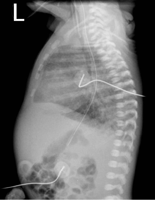
-The chest radiograph appearances of pneumonia are not specific, and frank lobar consolidation as seen in adults and older children is rare. More commonly, there is coarse opacity of one or more regions of the lung parenchyma. However, these appearances can also be seen with retained fetal lung fluid, meconium aspiration, aspiration of gastric contents, and pulmonary haemorrhage. Pleural effusions are not uncommon in infection, but again may be seen with other conditions.
-Group B Streptococcus can have an appearance similar to Respiratory Distress Syndrome (Surfactant Deficiency), although the granulation is typically more coarse.

-Transient Tachypnoea of the Newborn (TTN, also called Retained Fetal Lung Fluid or "Wet Lung") is a diagnosis of exclusion.
-Typical radiologic features are ill-defined but include:
1-Increased central vascular markings ("star-burst" appearance)
2-Hyperaeration
3-Evidence of interstitial and pleural fluid
4-Prominent interlobar fissures
5-Cardiomegaly
-Because the symptoms and radiological features are non-specific, infection should be considered in the differential diagnosis. Typically, respiratory symptoms resolve within the first 24-hours of life, but occasionally can persist longer.


- Respiratory Distress Syndrome (RDS) is a clinical diagnosis but one which is often interchanged with the terms Hyaline Membrane Disease (a pathological diagnosis) and Surfactant Deficiency (a term describing the typical appearances on radiographs of infants with RDS).


- The typical radiological features of Surfactant Deficiency are:
1-Small volume lungs
2-Homogenous "ground glass" opacity
3-Air bronchograms
-The differential diagnosis includes:
1-Pneumonia (particularly Group B Streptococcus)
2-Retained Fetal Lung Fluid/TTN
Findings
-This patient presents with an abnormal position of a nasogastric tube (NGT) overlying the left hemithorax.
-This patient demonstrates normally located chest tube and endotrachial tube (ETT).

Discussion
This patient suffered a rupture of the left hemidiaphragm secondary to a motor vehicle accident. The NGT is correctly placed in the stomach, but the stomach is now located in the left chest.


http://eradiology.bidmc.harvard.edu/Classics/item.aspx?section=Emergency+Radiology&labelpk=33f0adab-853f-4010-a0e2-0756b3f1eac5&pk=0513b7be-2271-4dc0-b365-1477cc2bf8ca
Findings
This patient presents with a sternomanubrial dislocation as demonstrated: There is posterior dislocation of the body of the sternum in relation to the manubrium of the sternum.

Discussion
Sternomanubrial dislocation is a rare injury, typically caused by high velocity trauma, e.g. MVA. There is a significant association with injury to the great vessels.


http://eradiology.bidmc.harvard.edu/Classics/item.aspx?section=Emergency+Radiology&labelpk=33f0adab-853f-4010-a0e2-0756b3f1eac5&pk=bc8b5dcf-8be2-4c84-a9ae-7cebe4673d01

Bullets and Shrapnel in the Chest on Plain Film and CT Scans

Findings
-This patient presents with two 9 mm bullets as outlined on plain film and CT scans: Bullet 1 is just deep to the ribs, bullet 2 is in the superficial soft tissues anterior to the sternum.

Discussion
-This patient was radiographed on a trauma backboard. When locating a foreign body on plain film, orthogonal views (views at 90 degrees to each other) are mandatory.


http://eradiology.bidmc.harvard.edu/Classics/item.aspx?section=Emergency+Radiology&labelpk=33f0adab-853f-4010-a0e2-0756b3f1eac5&pk=0b5aab3a-1116-4bad-9ddd-80ddbda0973c
Findings
-The CXR shows aberrant air secondary to the perforated esophagus; subcutaneous emphysema and pneumomediastinum.
-There is widening of the mediastinum and a left pleural effusion.
-The BaSw demonstrates extensive extravasation of contrast material through the lower esophageal perforation.
Discussion
Boerhaave’s Syndrome, or “spontaneous” esophageal perforation, usually presents with a linear tear through the lower esophageal wall secondary to vomiting against a closed glottis. The patients are frequently inebriated. Aberrant air is the usual radiologic presentation; pneumomediastinum occurs first. The air then tracks into the neck, producing deep cervical emphysema, which may rupture, producing subcutaneous emphysema. The pneumomediastinum may rupture into the pleural space causing a life threatening pneumothorax. Mediastinitis is the usual clinical presentation. Reported mortality estimate is approximately 35%, making it the most lethal perforation of the GI tract.

Tuesday, July 27, 2010

Findings
-Air is lucent (jet black) on plain film.
-There is streaky air in the subcutaneous tissue bilaterally.


http://eradiology.bidmc.harvard.edu/Classics/item.aspx?section=Emergency+Radiology&labelpk=33f0adab-853f-4010-a0e2-0756b3f1eac5&pk=01b624d2-41ea-4507-8a82-9cea3857bea1
Findings
-Aspiration may cause pneumonia in the dependent basilar segments.
-On lateral CXR, the normal spine should appear more lucent (darker) in the lower thorax.
-On lateral CXR, a positive lateral spine sign is said to be present if the lower thoracic vertebrae appear whiter.
-A positive lateral spine sign is frequently caused by basilar infiltrates.


http://eradiology.bidmc.harvard.edu/Classics/item.aspx?section=Emergency+Radiology&labelpk=33f0adab-853f-4010-a0e2-0756b3f1eac5&pk=6b5e5a96-e799-4206-b3fe-5e82667cd979
Findings
-The CXR demonstrates a widened mediastinum, indistinct aortic knob, and mediastinal shift to the right.
-The CT demonstrates an intimal flap in the descending aorta, a widened mediastinum secondary to periaortic hematoma, and a left-sided pleural effusion.
Discussion
-Aortic transection or rupture usually occurs as a result of high-impact blunt trauma e.g. MVA. 75%-90% result in immediate death. 18% of MVA-related deaths are secondary to aortic transection. Aortic transection is second only to head injury as the leading cause of injury-related death. Aortic transection must be distinguished from aortic dissection, which is a separation of the layers of the aortic wall, usually caused by a tear in the weakened intima. This patient demonstrates both with mediastinal hemorrhage and a Stanford Type B dissection.


http://eradiology.bidmc.harvard.edu/Classics/item.aspx?section=Emergency+Radiology&labelpk=33f0adab-853f-4010-a0e2-0756b3f1eac5&pk=6e919e0b-2a48-409b-a53c-32e51be6165a
Findings
-The CXR demonstrates a widened mediastinum.
-The CT demonstrates the intimal flap in the ascending and descending thoracic aorta.
Discussion
-Aortic dissection is the separation of the layers of the aortic wall, usually caused by a tear in the weakened intima . Aortic dissection is the most common fatal condition involving the aorta. The Stanford Classification has 2 types: Type A (60%) involves the ascending aorta. It may involve the arch or descending aorta to varying degrees. Type B (40%) involves the arch and/or descending aorta but not the ascending aorta. This is a Stanford type A aortic dissection, involving the origins of most of the aortic arch vessels. Notice that the aortic knob is still distinct and there is no mediastinal hematoma which differentiates this from an aortic transection.


http://eradiology.bidmc.harvard.edu/Classics/item.aspx?section=Emergency+Radiology&labelpk=33f0adab-853f-4010-a0e2-0756b3f1eac5&pk=f754f9fb-33f0-4a73-9afd-47845e2c601a