


 A
A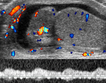 B
BClinically proved epididymitis in an 11-year-old boy. (a) Longitudinal US scan of the right hemiscrotum shows an enlarged hypoechoic epididymal head (E), reactive hydrocele (h), and thickening of the scrotal wall (*). m = mediastinum. (b) Color and pulsed-wave Doppler image shows increased vascularity in the epididymal head with a low-flow, low-resistance waveform pattern.


No comments:
Post a Comment