


 Lateral radiograph (A) of a 15-year-old boy shows a subperiosteal abscess that was aspirated and injected with contrast material. Anteroposterior (B) and lateral (C) radiographs of a 9-year-old child show contrast material in the subperiosteal area and within the medullary cavity after pus had been aspirated. The contrast material demonstrates the extent of the abscess.
Showing posts with label sinogram. Show all posts
Showing posts with label sinogram. Show all posts
Saturday, September 4, 2010
sinogram+sub periosteal abscess
Lateral radiograph (A) of a 15-year-old boy shows a subperiosteal abscess that was aspirated and injected with contrast material. Anteroposterior (B) and lateral (C) radiographs of a 9-year-old child show contrast material in the subperiosteal area and within the medullary cavity after pus had been aspirated. The contrast material demonstrates the extent of the abscess.
sinogram+osteomylitis
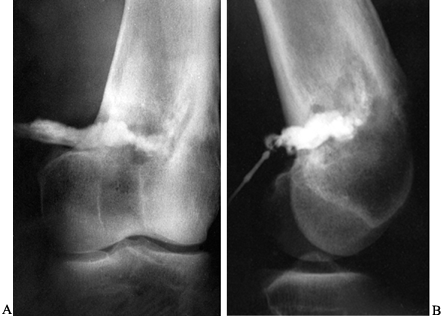 Anterior (A) and posterior (B) roentgenograms of a sinogram in a 21-year-old man with chronic osteomyelitis that tracks into the intramedullary cavity.
sinogram
What is sinogram?
It is a special X-ray procedure that is done with contrast dye to visualize any abnormal opening (sinus) in the body. The contrast is injected via a rubber catheter. Serial x-ray pictures are taken to show the extension of the fistula.
What kind of contrast medium used in this procedure?
A low osmolar contrast medium, LOCM 150.
How is it done?
Technique :
- A prelim film is taken to exclude the presence of radio-opaque foreign body.
- A fine catheter is then inserted into the orifice of the sinus.
- After a gauze pad has been firmly placed over the orifice to discourage reflux, the contrast medium is injected under fluoroscopic control.
- Spot films are taken as required including tangential views.
Below are sequence of films taken to investigate a sinus at right mid thigh.
1. Prelim : shows intramedullary fixation of right femur fracture. Site of fistula is located at mid thigh.

2. Right thigh AP : Dye is injected. There is a focal collection of contrast seen.

3. Lateral view : shows focal collection of contrast.

4. Right lower thigh AP : shows seepage of contrast seen into the intramuscular layers of the lateral aspect of the right thigh.


5. Right upper thigh AP : shows seepage of contrast seen into the intramuscular layers of the lateral aspect of the right thigh up to the level of hip joint and distally to the level of distal femur (just above the femoral condyles).


How is it reported?
This is a sample report of a sinogram case.NAME : ?
I/C : ?
SINOGRAM (01.04.2010)
Procedures:
Patient wound is cleaned. Sinus identified.
25 ml undiluted omipaque injected using 8F nasogastric tube.
Serial x-rays are taken.
Findings:
There is flow of contrast from the sinus into a focal collection measuring 3.5×4.5cm.
Seepage of contrast seen into the intramuscular layers of the lateral aspect of the right thigh.
Superiorly the contrast extends to the level of hip joint and distally to the level of distal femur (just above the femoral condyles).
There is no connection to the knee or hip joint.
CONCLUSION
No evidence of intra-articular extension of the right thigh abscess.
Radiologist 01.04.2010
Reference :
- A Guide to Radiological Procedures Stephen Chapman.


Subscribe to:
Comments (Atom)


