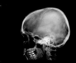
The skull image shows multiple round, punched-out lytic areas. A prominent lytic lesion is noted in the left femur on the next image and one in the right ischium on the third. The fourth image shows a small lesion in the posteriorlateral 5th rib. Note the lack of sclerotic margin.

No comments:
Post a Comment