Best Bath Store
All Natural Blend of 14 Herbs Gentle Cleanse- $20 Off Colonix Now! Use Promo Code 20J
Amberen - Breakthrough Menopause Treatment
728x90
Custom Search

Friday, May 28, 2010

dilated proximal right ureter.
at 1 cm caudal,right uerter passes behind IVC(arrow).

at 1 cm caudal,right ureter lies anterior to IVC,compare it
with left ureter which lie infront of left psoas muscle.

With CT, the proximal right ureter can be seen coursing medially behind and then anteriorly around the IVC so as to encircle it partially .


Reference:BiblioMed Textbook-Computed Body Tomography

superiorly,left IVC drain into right IVC through
retro aortic left renal vein.
open arrows=duplication of IVC.


Reference:BiblioMed Textbook-Computed Body Tomography
supra renal IVC is in its normal position.
at level of kidneys IVC crosses aorta via left renal vein(arrow).

here, we can find the calcified aorta on the right side
of infra renal IVC(inverted anatomical relation).

Reference:BiblioMed Textbook-Computed Body Tomography


Reference:BiblioMed Textbook-Computed Body Tomography


arrow head=showed adrenal gland related to duodenum
due abscense of IVC.
Arrow=azygous vein.
Arrow head=hemi azygous


at the level of tracheal bifurcation
arrow=azygous vein.
SVC=superior vena cava.


Reference:BiblioMed Textbook-Computed Body Tomography


Reference:BiblioMed Textbook-Computed Body Tomography

arrow=enlarged right gonadal vein in a patient

with right varicocele.


Reference:BiblioMed Textbook-Computed Body Tomography

The axial image demonstrates increased signal (arrow) within fat interposed between the lateral aspect of the patellar tendon and the lateral trochlear ridge. Mild lateral patellar subluxation is present as indicated by the off-midline positioning of the patellar tendon (arrowhead).
The sagittal image demonstrates increased signal (arrow) within the fat below the caudal margin of the patella as well as moderate patella alta (arrowhead). Note the high riding appearance of the patella.

Three normal anterior knee fat pads are present at the knee; the quadriceps (anterior suprapatellar) (red), the prefemoral (posterior suprapatellar or supratrochelar) (blue), and Hoffa (infrapatellar) (yellow) fat pads.


The infrapatellar fat pad is bordered by the inferior pole of the patella superiorly, the joint capsule and patellar tendon anteriorly, the proximal tibia and deep infrapatellar bursa inferiorly, and the synovium-lined joint cavity posteriorly. Thus, it is intracapsular but extrasynovial. It is tethered to the intercondylar notch superiorly by the infrapatellar synovial fold or infrapatellar plica. It also is attached directly to the anterior horns of the menisci inferiorly and to the periosteum of the tibia.


On MRI, a focal area of high signal or edema on STIR or T2 weighted sequences is present at the inferolateral aspect of the patellofemoral joint, specifically the lateral portion of the infrapatellar fat pad (D,E). Specifically, this is just below the inferior margin of the patella and anterior to the lateral trochlear ridge. A focal lobulated mass with signal characteristics of fluid or cystic change can sometimes be present in the lateral soft tissues of the knee between the lateral femoral condyle and the lateral retinaculum. Associated findings of lateral patellar subluxation (F) and/or patella alta are frequently present.


http://www.radsource.us/clinic/0809

Tuesday, May 25, 2010


Magnetic resonance imaging is as accurate as CT and arteriography in the detection of aortic dissection6. The MR diagnosis of an aortic dissection requires the demonstration of an intimal flap, which can be easily identified when both the true and false lumina appear as signal void areas on SE images .
Reference:BiblioMed Textbook-Computed Body Tomography
T1MRI image with GADOLINIUM revealed
aorta(A)surrounded by mural thrombus.
Reference:BiblioMed Textbook-Computed Body Tomography
short arrow=intimal calcification in anterior or posterior wall
long arrow=intact calcified intimal flap
look first to calcified intimal flap,if it is disrupted this indicate dissecting aneurysm,if it is
intact ,this will be calcification in the anterior or posterior wall of aortic aneurysm............................
Reference:BiblioMed Textbook-Computed Body Tomography
Dissection of the aorta usually originates in the thorax but sometimes extends into the abdomen. Its diagnosis is based on demonstration of an intimal flap with enhancement of both the true and false lumina after intravenous administration of contrast medium


Reference:BiblioMed Textbook-Computed Body Tomography
An infected atherosclerotic aortic aneurysm, likewise, can be demonstrated on CT as multiple air bubbles seen in the wall of the aneurysm.


Reference:BiblioMed Textbook-Computed Body Tomography
before contrast
after contrast

A mycotic aneurysm usually has a saccular, irregular contour, and it contains little or no mural calcification. It enhances to a degree similar to the adjacent normal appearing aorta .


Reference:BiblioMed Textbook-Computed Body Tomography

arrow=true aortic aneurysm.

arrow head=pseudo aortic aneurysm.


Reference:BiblioMed Textbook-Computed Body Tomography

Jumper's Knee refers to a spectrum of disorders that occur in patients with degeneration and/or tearing of the patellar tendon. It is one of the most common tendon abnormalities in athletically active individuals, and as the name implies, is most common in jumping athletes such as basketball and volleyball players. In the non-athlete, rheumatoid arthritis, the seronegative arthropathies, and treatment with exogenous steroids are known to predispose to this condition.
fat-suppressed proton density-weighted sagittal images in a basketball player who sustained an acute injury reveal marked edema about the patellar tendon with a fluid filled gap (arrows) at the central tendon, compatible with a complete rupture. The retracted proximal and distal ends (arrowheads) are markedly thickened, indicating severe tendinosis. The vast majority of patellar tendon ruptures occur in patients with pre-existing patellar tendinosis.
A proton density-weighted axial view demonstrates the normal semilunar appearance of the patellar tendon, with a convex anterior border (arrow) and well-defined posterior rim.
T1-weighted sagittal image in a patient with a normal patellar tendon reveals a homogeneously low signal intensity appearance to the tendon (arrows), which appears symmetrical in appearance throughout its course.

http://www.radsource.us/clinic/0612
The fat-suppressed proton density weighted sagittal view confirms the abnormally thickened and edematous tendon (arrow). Mild adjacent edema is seen within the surrounding subcutaneous and infrapatellar fat, and small interstitial splits (arrowheads) are present within the proximal tendon.
The T1-weighted sagittal images reveals marked thickening and increased signal intensity (arrow) within the patellar tendon.

http://www.radsource.us/clinic/0612
A=aortic aneurysm.
short arrows=hyperdense crescent seen
suggesting early rupture.
Reference:BiblioMed Textbook-Computed Body Tomography
A=aortic aneurysm.
short black arrow=mural thrombus.
H=hyperdense STREAKY SHAPED hematoma related to psoas muscle(P).
Reference:BiblioMed Textbook-Computed Body Tomography
aorta calcified and markedly dilated.
bilateral dilatation of both proximal
common iliac arteries.


Reference:BiblioMed Textbook-Computed Body Tomography

contrast enhanced aorta showed dilated aorta with 7cm diameter
white arrow=rim of intact intimal calcification.
black arrow=cresentic mural thrombus.
I=IVC
D=duodenum.
Reference:BiblioMed Textbook-Computed Body Tomography

arrow=atheroma in aorta,clearly differentiated
from contrast enhanced aorta.
Reference:BiblioMed Textbook-Computed Body Tomography
mediastinal hematoma surrounding esophagus(long arrow)
and aorta (short arrow).
long black arrow=small sized aorta(7mm) due to shock
following vehicle accident.
long white arrow=retro peritoneal hemorrhage.
short white arrow=intra peritoneal hemorrhage.

Reference:BiblioMed Textbook-Computed Body Tomography

Monday, May 24, 2010

Piriformis syndrome is an uncommon cause of buttock and hip pain due to entrapment of the sciatic nerve by the piriformis muscle at the greater sciatic notch.
The coronal T1-weighted images demonstrate enlargement of the right piriformis muscle (arrow).
The corresponding axial T2-weighted image confirms enlargement of the right piriformis muscle (arrow).

A coronal inversion recovery weighted image demonstrated no abnormal signal intensity within either piriformis muscle (arrows).


Sunday, May 23, 2010

uncommon disease.
known as recurrent pyogenic cholangitis or oriental cholangitis.


There is intrahepatic lithiasis with focal diatation.
There is focal dilatation of the bile ducts in the left lobe with stones.
The left lobe is the most common location of the disease due to the delayed drainage of the left system.
Klatskin Tumor
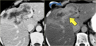
Intrahepatic cholangiocarcinoma. Capsular retraction and the late enhancement
Early and late phase enhancement of a intrahepatic cholangiocarcinoma

The key findings to look for are:
•Delayed enhancement
•Peripheral biliary dilatation
•Capsular contraction


http://www.radiologyassistant.nl/en/49e17de25294d#a49e4e5125f1b4
On the CT the liver looks quite normal.
However if you look at the common bile duct in the pancreatic head you will notice the soft tissue density.
The differential diagnosis would include an impacted stone or cholangiocarcinoma, but since this patient had no obstruction, it was thought to be the result of bile wall thickening.
The gallbladder wall is also thickened.
arrow=irregular thickening of common bile
duct walls
cholangio carcinoma within choledochal cyst.