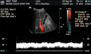
 Changing the wall filter. (a) Color duplex US image obtained with a high wall filter setting shows loss of the low-velocity-flow component of the spectral waveform immediately above the baseline. Higher-velocity flow is well depicted, and accurate flow quantification can still occur. In the evaluation of the liver vasculature, this is likely to become relevant only when flow velocity is very low and falls within the range of velocities that are filtered out. (b) Color duplex US image demonstrates how the spectral waveform progressively fills in toward the baseline as the wall filter is sequentially reduced from high (left arrow) to medium (middle arrow) to low (right arrow).


No comments:
Post a Comment