


 Enhancing right upper lobe collapse with sharp margins delineated laterally with minor fissure and posteriorly with major fissure.
 Evidence of volume loss in the right hemithorax.
white arrows:lesser fissure.
open arrow:greater fissure.
 obstruction in the lobar bronchus is demonstrated(black arrow).
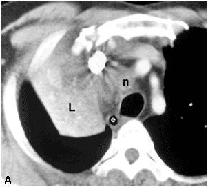
 n=mediastinal lymphadenopathy
 obstruction in the lobar bronchus is demonstrated(black arrow).
 n=mediastinal lymphadenopathy
black arrow head=compressed superior vena cava
white arrows=collapsed right upper lobe
black arrow=obstructed upper lobe bronchus.


No comments:
Post a Comment