


 portal hypertension&varices
 portal hypertension &varices
 ascitis
 Omental cake refers to infiltration of the omental fat by material of soft-tissue density. The most common causes are metastases from ovary, stomach or colon. Tuberculous peritonitis may also give this appearance. Masses on the peritoneal surfaces may also be present. Malignant ascites may be present.
A) B)
B)
C) D)
D)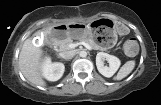 Images in a 66-year-old woman with a right upper quadrant abscess that occurred after cholecystectomy. (a) Transverse contrast-enhanced supine CT image shows abscess (arrow) in the gallbladder fossa. (b) Sagittal US image of the abscess obtained immediately prior to US-guided drainage shows multiple septations (arrow). (c) Transverse contrast-enhanced supine CT image shows residual abscess collection (arrow) identified next to the drainage catheter. (d) Transverse contrast-enhanced supine CT image obtained following intracavitary administration of 4 mg of tPA in 50 mL of 0.9% saline twice daily for 3 days shows no residual collection. The catheter was then removed, and there was no recurrence of the abscess cavity.

B)C)
D) Images in a 66-year-old woman with a right upper quadrant abscess that occurred after cholecystectomy. (a) Transverse contrast-enhanced supine CT image shows abscess (arrow) in the gallbladder fossa. (b) Sagittal US image of the abscess obtained immediately prior to US-guided drainage shows multiple septations (arrow). (c) Transverse contrast-enhanced supine CT image shows residual abscess collection (arrow) identified next to the drainage catheter. (d) Transverse contrast-enhanced supine CT image obtained following intracavitary administration of 4 mg of tPA in 50 mL of 0.9% saline twice daily for 3 days shows no residual collection. The catheter was then removed, and there was no recurrence of the abscess cavity. 
sub-phrenic abscess
 intra-abdominal abscess


No comments:
Post a Comment