Best Bath Store
All Natural Blend of 14 Herbs Gentle Cleanse- $20 Off Colonix Now! Use Promo Code 20J
Amberen - Breakthrough Menopause Treatment
728x90
Custom Search

Monday, June 14, 2010

(WHO grade 111)

Anaplastic astrocytoma. Axial T2-weighted and enhanced Tl -weighted images demonstrate a large right temporal mass with prominent enhancement and extensive surrounding infiltration. Differential diagnosis includes lymphoma. (Click to magnify figure)

http://yassermetwally.wordpress.com/brain-tumors/adult-brain-tumors/

(WHO grade 11)

World Health Organization (WHO)Grade II infiltrating astrocytoma(low grade). Axial T2-weighted, FLAIR, and enhanced Tl -weighted images. There is a high signal intensity mass in the left frontal lobe. No significant edema or enhancement is identified. (Click to magnify figure)

acute stage

Acute hemorrhage is hyperdense (white) on CT scan
A
B

C
Cerebral infarction is hypodense (black) on CT scan. With progression of time (from A to C) the infarction gets more hypodense, more well defined and the mass effect gradually decreases with time due to gradual reduction of brain edema because the blood brain barrier is once again sealed. The initial hypodensity in acute infarction is due to edema (A) while the the ultimate hypodensity in old infarction (C) is due to astrogliosis with widened fluid filled extracellular spaces (microcavitations and macrocavitations). During the evolution of the infarction the edema and the swelling decreases and the infarction boundary becomes better defined, and the infarcted area becomes more hypodense.

Sunday, June 13, 2010

types


definition

Cerebral palsy (CP) is an umbrella term encompassing a group of non-progressive, non-contagious conditions that cause physical disability in human development.
Cerebral refers to the cerebrum, which is the affected area of the brain (although the disorder most likely involves connections between the cortex and other parts of the brain such as the cerebellum), and palsy refers to disorder of movement. CP is caused by damage to the motor control centers of the young developing brain and can occur during pregnancy (about 75 percent), during childbirth (about 5 percent) or after birth (about 15 percent) up to about age three.
It is a non-progressive disorder, meaning the brain damage does not worsen, but secondary orthopedic difficulties are common. There is no known cure for CP. Medical intervention is limited to the treatment and prevention of complications arising from CP's effects.
Magnetic resonance imaging showed cystic encephalomalacia of LMCA territory, suggesting prenatal insult,

http://www.google.com.eg/imgres?imgurl=http://www.sahha.gov.mt/showdoc.aspx%3Fid%3D456%26filesource%3D4%26file%3DFIG01.JPG&imgrefurl=http://www.sahha.gov.mt/pages.aspx%3Fpage%3D456&usg=__9iZwz9QeCdEhkiEsasxisd4g9Nc=&h=434&w=354&sz=22&hl=en&start=12&itbs=1&tbnid=wCn7XSbrTvNSbM:&tbnh=126&tbnw=103&prev=/images%3Fq%3Dcerebral%2Bpalsy,mri%26hl%3Den%26tbs%3Disch:1
- The most frequent MRI finding was a focal thinning at the isthmus of the corpus callosum
-The second most frequent MRI finding was ventricular asymmetry in the axial and coronal plane
-The next most frequent MRI finding was leukomalacia associated or not with other lesions in the periventricular region . These lesions presented hyperintensity on T2-weighted and FLAIR acquisitions.
-Another frequent finding identified in 12 (16%) of the L children was gliosis, defined as the presence of a round or oval white matter lesions that presented isointensity on T1-weighted images and hyperintensity on T2-weighted and FLAIR images.
-Arachnoid cysts


http://www.google.com.eg/imgres?imgurl=http://www.scielo.br/img/revistas/anp/v64n2a/a03fig02.gif&imgrefurl=http://www.scielo.br/scielo.php%3Fscript%3Dsci_arttext%26pid%3DS0004-282X2006000200003&usg=__aMFhBUWip9a_tq7814OL_mEvhwk=&h=607&w=440&sz=174&hl=en&start=4&itbs=1&tbnid=KWsxgXUebWQBkM:&tbnh=136&tbnw=99&prev=/images%3Fq%3Dcerebral%2Bpalsy,mri%26hl%3Den%26tbs%3Disch:1
Magnetic resonance imaging (MRI) scan of a 9-day-old girl who was born full-term and had a perinatal hypoxic-ischemic event. Examination of the patient at 1 year revealed findings consistent with a mixed quadriparetic cerebral palsy notable for dystonia and spasticity. Severe hypoxic-ischemic injury to the medial aspect of the cerebellar hemispheres, medial temporal lobes, bilateral thalami, and bilateral corona radiata is observed.

emedicine.medscape.com/article/310740-overview

Magnetic resonance imaging (MRI) scan of a 1-year-old boy who was born at gestational week 27. Clinical examination is consistent with spastic diplegic cerebral palsy. Pseudocolpocephaly and decreased volume of the white matter posteriorly are consistent with periventricular leukomalacia. Evidence of diffuse polymicrogyria and thinning of the corpus callosum is noted.
Magnetic resonance imaging (MRI) scan of a 16-month-old boy who was born at term but had an anoxic event at delivery. Examination findings are consistent with a spastic quadriplegic cerebral palsy with asymmetry (more prominent right-sided deficits). Cystic encephalomalacia in the left temporal and parietal regions, delayed myelination, decreased white matter volume, and enlarged ventricles can be seen. These findings are most likely the sequelae of a neonatal insult (eg, periventricular leukomalacia with a superimposed, left-sided cerebral infarct).

http://www.google.com.eg/imgres?imgurl=http://img.medscape.com/pi/emed/ckb/rehabilitation/305143-310739-310740-1645210.jpg&imgrefurl=http://emedicine.medscape.com/article/310740-overview&h=504&w=424&sz=47&tbnid=EyT-AJf9ko_pVM:&tbnh=245&tbnw=206&prev=/images%3Fq%3Dcerebral%2Bpalsy,mri&usg=__60jEl9ayucZ8DlCfm5ni6xabPM4=&sa=X&ei=fDIVTO2wCcj84AaD0onMCA&ved=0CBoQ9QEwAA
Pancreatic involvement in acute lymphoblastic leukaemia is rare [1, 2, 3] and obstructive jaundice secondary to a pancreatic mass as a primary presentation of acute lymphoblastic leukaemia has not been reported in the surgical literature. Acute lymphoblastic leukaemia typically presents with symptoms of bone marrow failure such as fatigue, lethargy, infections, bruising or bleeding. Approximately half the patients will have lymphadenopathy, splenomegaly or hepatomegaly at presentation. Full blood count may reveal cytopenias or (as in this case) a raised white cell count due to circulating blast cells. Although long term survival in adults is less good than children, acute lymphoblastic leukaemia is an important diagnosis to make because it is highly chemo-sensitive, with 91% of adults achieving complete remission following induction therapy in the recent UK ALL XII trial [4]. If suspected, a haematological referral is required since the diagnostic procedure of choice is a bone marrow aspirate and trephine.

http://www.blogger.com/www.joplink.net/prev/201001/19.html
A four-year-old boy with sacrococcygeal endodermal sinus tumour. Blood culture grew Peiciliomyces lilacinus. (a) CT lungs showed tiny nodular lesions, presumed involvement of lungs by fungus. (b) After two weeks of anti-fungal therapy, CT showed the lung lesions had increased in size and number. (c) CT lungs after two months of continuous anti-fungal therapy showed more and larger lesions, which were likely to be metastases.

http://www.blogger.com/www.biij.org/2006/2/e21/
Fungal abscesses in intrabdominal organs. (a) A two-year-old girl with leukaemia and Aspergillus sepsis. CT showed multiple hypodense lesions in both kidneys. (b) A six-year-old girl with leukemia and presumed fungal sepsis. CT revealed multiple hypodense lesions in the liver and spleen. Fungal elements were isolated from liver biopsy, but the species could not be identified. (c) A three-year-old boy with leukemia with presumed fungal sepsis. CT abdomen showed multiple hypodense splenic lesions No fungi were isolated from biopsy.

http://www.blogger.com/www.biij.org/2006/2/e21/
Fungal sinusitis of a four-year-old girl with myeloid leukaemia. CT of the sinuses revealed opacification of the maxillary sinuses with bone destruction of the medial wall. Mucor species was isolated from maxillary sinus washout.

http://www.blogger.com/www.biij.org/2006/2/e21/
A 10-year-old girl with leukemia and biopsy proven Aspergillus infection of the lung. She later developed a mycotic aneurysm in the chest. (a) CT showed a cavity with a “crescent sign” in keeping with an Aspergilloma. The lesion was excised. (b) Chest radiograph two months later showed a round opacity in the right lung. (c) CT revealed an aneurysm of the lower lobe branch of the right pulmonary artery.

http://www.blogger.com/www.biij.org/2006/2/e21/
An 18-month-old girl with leukaemia and Aspergillus isolated from the lung. (a) Chest radiograph showed a round opacity behind the heart. (b) CT revealed a cavitating nodule in the left lower lobe.
Neck CT scan demonstrating large thyroid lymphoma
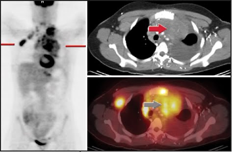
Marked FDG uptake throughout the mediastinum and in the right axilla/supraclavicular area corresponding to bulky adenopathy on the CT portion of the exam compatible with malignancy.
Chemotherapy (CHOP). Follow-up PET/CT ordered following 1 cycle.
Complete resolution of abnormal FDG activity compatible with a good response to therapy. Focal apparent FDG activity in the left supraclavicular area was not present on the uncorrected images compatible with an attenuation correction artifact. Bulky adenopathy is still present, but no increased FDG activity is present.
Discussion:
This case demonstrates the power of PET/CT to assess response to therapy soon after initiation. The strength of the modality is in the ability to assess an early response to therapy by assessing the metabolic changes. As shown in the second set of images, there is still considerable soft tissue abnormality present, but no increased FDG activity. Evidence suggests that for non-Hodgkin’s lymphoma, patients are to be categorized as responders (better overall survival) only if there is minimal or no residual FDG activity on follow up exams after therapy initiation. The metabolic changes can be assessed after one cycle of chemotherapy, whereas the soft tissue component will take much longer to regress and may remain indefinitely.

Esophageal involvement by mediastinal lymphoma. CT scan in a patient with large cell lymphoma of the mediastinum shows extensive mediastinal adenopathy compressing the esophagus (arrowhead) and superior vena cava (arrow). (Courtesy of Duane G. Mezwa, MD, Royal Oak, MI.)
CT non contrast
ADC MAP

DWI


Axial flair


MR perfusion CBV map
findings:Multiple enhancing mass lesions with increased choline and restricted diffusion and minimally decreased perfusion.
D.D.:Metastases, abscesses, lymphoma, demyelination
Discusion:Lymphoma with high cellularity may show restricted diffusion and iso or slightly decreased perfusion.

CT showed lymphadenopathy above and below the diaphragm and a lesion in L-3 vertebra consistent with a compressed or pathological fracture. The final diagnosis of lymphoma was made after biopsy of enlarged right inguinal lymph nodes and by bone marrow biopsy. There are areas of increased pathological activity in the left supra and infraclavicular region, the upper and mid-mediastinum, the right pulmonary hilus, D-8, D-12, L-2, L-3, L-4 vertebrae and the left inguinal region.

http://www.blogger.com/www.gehealthcare.com/.../products/lymphoma.html
Contrast-enhanced helical CT scan demonstrates a markedly enlarged, conglomerate nodal mass with homogeneous attenuation enveloping the retroperitoneum. Lymphoma was the most likely diagnosis, but biopsy revealed adenocarcinoma.

radiographics.rsna.org/.../1/197/F38.expansion

orbital lymphoma


This is 68 year old man with bilateral propotosis and lymphadenopathy. CT images are characterstic of orbital lymphoma.
PET/CT for staging of Hodgkin's lymphoma. CT showed involvement only in right neck. PET/CT (A: coronal views; B: transverse views; MIP = maximum-intensity projection) showed that normal-size (9-mm) upper mediastinal lymph node was clearly metabolically active, changing stage from I to II. This finding is relevant if consolidative radiation after chemotherapy is planned. Incidental normal scalene muscle uptake was noted on coronal PET.

http://jnm.snmjournals.org/cgi/content-nw/full/48/1_suppl/19S/FIG1
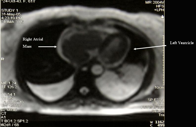
Primary right atrial lymphoma. Magnetic Resonance Imaging # Description : Magnetic Resonance Imaging of primary right atrial lymphoma
Primary right atrial lymphoma. CT scan of jugular veins # Description : Contrast CT scan showing extension of the primary right atrial lymphoma through the superior vena cava up to the right jugular vein that is occluded while the contra-lateral jugular vein is opened.


Figure 1. 55-year-old man with recurrent mantle cell lymphoma. CT shows symmetric circumferential thickening of the distal trachea (arrows).
Figure 2. 55-year-old man with recurrent mantle cell lymphoma. Axial PET/CT shows intense activity in the suspected area around the distal trachea.


Figure 3. 55-year-old man with recurrent mantle cell lymphoma. Full body coronal reformatted PET scan shows abnormal intense tracheal uptake and other areas of expected uptake.


The baseline CT's showed lypmhadenopathy in the left inguinal node and the left iliac fossa. The PET Scan showed increased metabolism in both of these lesions, however the Gallium scan only showed uptake in the inguinal lesion. The Bone scan was normal.
This abdominal CT scan shows tumor masses (malignant lymphomas) in the area behind the peritoneal cavity (retroperitoneal space).

cylindrical or tubular type

signet appearance due to dilated bronchus in near to
pulmonary artery.
In normal individuals, the adjacent pulmonary artery is the same size, or minimally smaller, than the bronchus. A bronchoarterial ratio >1.2 is indicative of abnormal bronchial dilatation. Substantial dilatation of the bronchus and smooth mural thickening results in a signet ring configuration
Reference:BiblioMed Textbook-Computed Body Tomography

cylindrical or tubular type

arrowhead=bronchial wall thickening in transverse section.
short arrow= in longitudinal section.
Normal bronchi are not visible on CT in the most peripheral areas of the lung, within 2 cm of the costal or paravertebral pleura. The cardinal sign of bronchiectasis on HRCT is dilatation of the bronchus, which usually is accompanied by bronchial wall thickening (internal diameter <80%>1.2 is indicative of abnormal bronchial dilatation.

Reference:BiblioMed Textbook-Computed Body Tomography

Saturday, June 12, 2010

ESSENTIALS
-Many patients with inflammatory or infectious types of colitis present with abdominal pain, and multidetector CT is often used as the initial diagnostic test.
-CT can accurately depict the bowel wall and help determine the presence and extent of intramural disease, its possible extraluminal extension, and involvement of distant sites.
-Many imaging features or patterns are helpful in distinguishing the various types of colitis, including degree of wall thickening, extent and location of disease, extraluminal manifestations, and types of complication.
-While imaging features can help narrow the differential diagnosis or suggest a specific diagnosis, the final diagnosis is often based on clinical and laboratory data and colonoscopic and biopsy findings.

radiology.rsna.org/content/240/3/623.full
Transverse CT image in a 67-year-old woman with acute right upper quadrant pain due to omental infarction shows ill-defined, ovoid, fatty mass (arrows) of increased attenuation centered in the greater omentum.

radiology.rsna.org/content/240/3/623.full
Transverse CT image in a 34-year-old man with acute right lower quadrant pain caused by appendicitis with perforation. Appendiceal lumen (short black arrow) is enlarged and shows wall enhancement. Mesenteric stranding (white arrow), free intraperitoneal air (arrowhead), and thickened terminal ileum (long black arrow) due to contiguous inflammation are seen.

radiology.rsna.org/content/240/3/623.full
Transverse CT image in a 21-year-old man with right lower quadrant pain due to appendicitis shows an appendicolith (arrowhead) separated from cecal lumen by cecal wall thickening (cecal bar sign) (black arrow). Dilatation of inflamed appendix with air and debris (white arrow) is demonstrated.

radiology.rsna.org/content/240/3/623.full

Transverse CT image in a 57-year-old woman with diverticulitis of sigmoid colon shows wall thickening (large arrows) in sigmoid colon and scattered diverticula. An inflamed diverticulum (arrowheads) and mesenteric fluid (small arrow) also are seen.

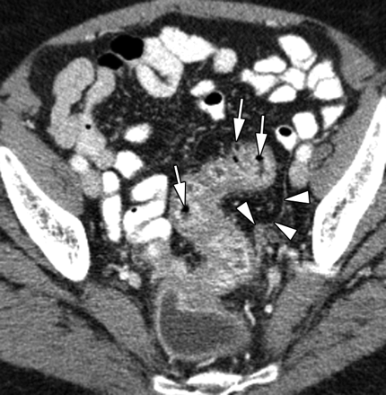
Transverse CT image in a 65-year-old woman with early diverticulitis who had left lower quadrant pain and leukocytosis reveals wall thickening and multiple small diverticula (arrows) in a long segment of the sigmoid colon. Fascial thickening (arrowheads) along left pelvic side wall indicates mild diverticulitis.
a
b
Ischemic colitis in an 81-year-old woman with myocardial infarction. (a) Anteroposterior CT scout view shows air (arrows) in wall of right colon and small- and large-bowel dilatation. (b) Transverse CT image demonstrates air (arrowheads) in wall of right colon, with lack of wall enhancement and pericolonic stranding indicative of infarction.