Best Bath Store
All Natural Blend of 14 Herbs Gentle Cleanse- $20 Off Colonix Now! Use Promo Code 20J
Amberen - Breakthrough Menopause Treatment
728x90
Custom Search

Friday, July 2, 2010

Hysterosalpingography (left image): Severe dilatation of the tubes following a short intact section (black arrows) is visible. The contrast material cannot get into the peritoneal cavity. Operation is not possible because the intact part of the tube is short.
Right image: Significant dilatation of only the distal part of the tubes (arrows) is visible, therefore the abnormal part can be resected.


http://http://www.google.com.eg/imgres?imgurl=http://www.szote.u-szeged.hu/radio/emlo/emlo9c.gif&imgrefurl=http://www.szote.u-szeged.hu/radio/emlo/aemlo9c.htm&usg=__MS-RJu8NFOZKRjpw2vKugRuLzhM=&h=400&w=654&sz=187&hl=en&start=6&itbs=1&tbnid=vZeWr6VdjuIBhM:&tbnh=84&tbnw=138&prev=/images%3Fq%3Dhysterosalpingography%26hl%3Den%26sa%3DG%26gbv%3D2%26tbs%3Disch:1

normal case

Hysterosalpingography: Normal anatomical structures. The contrast material is injected through a Schultze-device placed into the cervix. The contrast material outlines the uterine cavity and the thin Fallopian tubes opening into the peritoneal cavity.

Wednesday, June 30, 2010

FIGO classification of cervical carcinoma

The major categories of the FIGO classification are as follows:
Stage 0 – Carcinoma in situ
Stage I – Invasive carcinoma that is strictly confined to the cervix
Stage II – Locoregional spread of the cancer beyond the uterus but not to the pelvic sidewall or the lower third of the vagina
Stage III – Cancerous spread to the pelvic sidewall or the lower third of the vagina, and/or hydronephrosis or a nonfunctioning kidney that is incident to invasion of the ureter
Stage IV – Cancerous spread beyond the true pelvis or into the mucosa of the bladder or rectum
The FIGO stages are further categorized as follows:
Stage Ia cervical carcinoma – Preclinical invasive carcinoma that can be diagnosed only by means of microscopy
Stage Ib cervical carcinoma – A clinically visible lesion that is confined to the cervix uteri
Stage Ib1 – The primary tumor is not greater than 4.0 cm in diameter.
Stage Ib2 – The primary tumor is greater than 4.0 cm in diameter.
Stage IIa cervical carcinoma – Spread into the upper two thirds of the vagina without parametrial invasion
Stage IIb cervical carcinoma – Extension into the parametrium but not into the pelvic sidewall
Stage IIIa cervical carcinoma – Extension into lower one third of the vagina, without spread to the pelvic sidewall
Stage IIIb cervical carcinoma – Extension into the pelvic sidewall and/or causes a nonfunctioning kidney or hydronephrosis due to invasion of the ureter
Stage IVa cervical carcinoma – Extension of the tumor into the mucosa of the bladder or rectum
Stage IVb cervical carcinoma – Spread of the tumor beyond the true pelvis and/or by metastasis into distant organs
The strict FIGO clinical staging guidelines do not include the status of the lymph nodes, although the presence of metastatic adenopathy is an important factor in treatment planning and in the prognosis.
Extended clinical staging with cross-sectional imaging (CT scanning and/or MRI) includes the status of the lymph nodes in the assessment of the extent of the disease. The detection of enlarged pelvic lymph nodes is considered equivalent to pelvic sidewall tumor extension (stage III), and the detection of enlarged lymph nodes in the para-aortic, paracaval, or inguinal regions is considered extrapelvic tumor spread (stage IV).
The major limitations of the FIGO clinical staging system are encountered in the estimation of the size of the primary tumor, particularly when the tumor is endocervical. The size of the tumor is significant because, in each stage, the incidence of lymph node metastases increases and the prognosis deteriorates with increased volume of the primary tumor.
Other limitations occur in the evaluation of tumor extension into the parametrium and pelvic sidewalls and in the detection of metastatic lymphadenopathy or distant metastasis.Extended clinical staging with cross-sectional imaging (CT scanning and/or MRI) and surgicopathologic staging, including pelvic and abdominal retroperitoneal lymphadenectomy, provide additional diagnostic value.
Each has been proven to be superior to the conventional FIGO clinical staging system in determining the full extent of the tumor spread. However, once the clinical stage is assigned on the basis of the clinical pretreatment workup results (in compliance with the FIGO guidelines), the stage should not be altered as a result of subsequent findings. Instead, any additional information that is revealed by cross-sectional imaging or surgery is primarily used for planning treatment regimens, and they should not be used to revise the assigned clinical stage.


http://emedicine.medscape.com/article/402329-media
Human papillomavirus (HPV) is now recognized as the most important causative agent in cervical carcinogenesis at the molecular level, although HPV may not induce many of the identified molecular alterations.11,12 As many as 5% of cervical cancers may not be associated with HPV.13,14 First intercourse at an early age, sexual promiscuity, high parity, race, and low socioeconomic status are presently thought to increase the risk for cervical cancer because these factors are linked to sexual behavior that increases the likelihood of exposure to HPV and/or because they are cofactors that modify the risk in women who are infected with HPV. Tobacco smoking is also a significant independent risk factor.

http:// emedicine.medscape.com/article/402329-media
spread of cervical carcinoma
1-The main pathways for the spread of invasive cervical cancer consist of the following: (1) microscopic spread into the vaginal mucosa beyond a visible or palpable tumor; (2) extension into the endometrium or myometrium of the corpus; (3) direct extension into the parametrium and, in advanced stages, into the adjacent structures; and (4) spread into the regional pelvic lymph nodes and, potentially, into the retroperitoneal, inguinal, or thoracic lymph nodes.
2-The extrauterine spread of cervical cancer occurs primarily by means of direct extension and lymphatic invasion that initially affects the contiguous tissues in the region of the laterally positioned cardinal ligament. Later, the tumor may involve the anterior or posterior parametrium, which are separated from the cervix by the pubovesicocervical fascia. The lateral paracervical and parametrial regions are more vulnerable to tumor invasion than the anterior and posterior parametrium because of the lack of a protective fascial covering at the lateral regions and because of the natural lymphatic drainage through the lateral paracervical tissues into the cardinal ligaments.
3-The local spread of cervical cancer may progress through the parametrium to involve the ureters and, eventually, the pelvic sidewalls. In some patients, the sciatic plexus is involved. Hydronephrosis, pyelonephritis, and renal failure are common complications of progressive disease. Involvement of the urinary bladder and rectum can occur in advanced cases because of direct tumor extension or subsequent to invasion of the vesicouterine or uterosacral ligaments, respectively. A vesicovaginal fistula or rectovaginal fistula may or may not develop. A tumor that extends through the posterior aspect of the cervix or corpus infrequently leads to intraperitoneal spread. Adnexal metastases are uncommon intheearlystages of the disease.
4-Lymphatic tumor spread usually occurs in a fairly orderly pattern or sequence that first involves the regional paracervical and parametrial lymph nodes and then the internal and external iliac lymph nodes. This may then be followed by spread to the common iliac nodes, the para-aortic nodes, and eventually the supraclavicular nodes via the thoracic duct. Metastasis to the para-aortic lymph nodes without involvement of pelvic lymph nodes is unusual.
5-Hematogenous tumor spread may be a result of a lymphatic venous anastomosis or direct venous invasion. The most common sites of hematogenous metastases are the lungs, bones, and liver.

http://emedicine.medscape.com/article/402329-media

Computed tomography (CT) scan of clinical stage IIb cervical carcinoma (same patient in Images 4-5 in Multimedia). The parametrial invasion is depicted with CT scanning as loss of definition of the cervical contours, accompanied by increased attenuation and prominent soft-tissue stranding in the parametrial fat. Parametritis can result in similar findings. The cervix shows ill-defined hypoattenuation, but the tumor is not clearly delineated. In addition, a subserosal leiomyoma protrudes from the left side of the uterus.
Computed tomography scan of a large, lobulated mass that is replacing the cervix and showing nonuniform hypoattenuation. The air and fluid in the center of the mass are consistent with tumor necrosis and a complicating infection (the patient had purulent discharge). The central hypoattenuation in the uterine corpus is suggestive of minimal fluid in the cavity.

http:// emedicine.medscape.com/article/402329-media

Computed tomography scan through the upper uterus (same patient in Images 1-2 in Multimedia). This image shows fluid that markedly distends the endometrial cavity secondary to obstruction of the endocervical canal by cervical cancer. A small submucosal leiomyoma projects into the right anterior aspect of the endometrial cavity and has minute calcifications.

http://%20emedicine.medscape.com/article/402329-media


Computed tomography (CT) scan of parametrial and rectal invasion by cervical carcinoma (same patient in Images 10-11 in Multimedia). There is loss of definition of the cervical contours, accompanied by a masslike soft tissue that replaces the parametrial fat on the right and that extends into the anterior and right-sided rectal walls
This computed tomography scan demonstrates a markedly enlarged lymph node at the left pelvic sidewall, a finding that is consistent with pelvic lymph node metastasis, which is indicative of stage IIIb disease. The cystic consistency is not unusual for metastatic cervical carcinoma. The primary tumor is well depicted as a hypoattenuating, circumscribed mass. A cyst is present in the anteriorly located left ovary.

http://emedicine.medscape.com/article/402329-media
This computed tomography scan demonstrates a cervical tumor directly extending into the posterior wall of the bladder and into the left pelvic sidewall. Extension into the pelvic sidewall is a feature of stage IIIb disease, whereas involvement of the bladder wall is a feature of stage IVa disease.

http:// emedicine.medscape.com/article/402329-media


Computed tomography (CT) scan of stage IVb cervical carcinoma (same patient in Images 14-17 in Multimedia). There is the presence of borderline enlarged para-aortic and pelvic lymph nodes, presumably secondary to metastasis; left hydronephrosis and hydroureter to the level of the large cervical tumor; and direct intraluminal extension of the tumor into the bladder. The borderline enlarged left para-aortic lymph node is consistent with stage IVb cervical carcinoma.

Sagittal T2-weighted magnetic resonance image (MRI) of a large cervical tumor extending through the entire cervical stroma and extensively involving the uterine corpus, vagina, bladder wall, and posterior urethral region . The invasion of the bladder wall is a feature of stage IVa disease. Courtesy of Kaori Togashi, MD, Hitachi Medical Corporation, Chair of Department of Diagnostic and Interventional Imageology, Kyoto University, Japan.
Axial T2-weighted magnetic resonance image (MRI) of a large cervical tumor with full-thickness stromal invasion causing complete loss of the hypointense stromal stripe or ring (same patient in Images 22-23 in Multimedia). Also depicted is invasion of the parametrium and the posterior bladder wall; this finding is indicative of stage IVa disease. Courtesy of Kaori Togashi, MD, Hitachi Medical Corporation, Chair of Department of Diagnostic and Interventional Imageology, Kyoto University, Japan.

http:// emedicine.medscape.com/article/402329-media
This sagittal transabdominal sonogram shows a circumscribed hypoechoic tumor in the posterior aspect of the cervix

http://emedicine.medscape.com/article/402329-media
This transverse transvaginal sonogram shows a circumscribed hypoechoic tumor in the left posterior aspect of the cervix

http://emedicine.medscape.com/article/402329-media

This sagittal transvaginal color Doppler sonogram shows prominent vascular flow in the cervical tumor .

ultrasound technique

First, the entire abdomen is examined with a 4- to 5-MHz curved sector scanner. There are a number of other etiologies of abdominal or RLQ pain, ranging from mesenteric adenitis, to obstructive uropathy, to ovarian torsion. Thus, an examination of the entire abdomen is clearly warranted. In women, one must examine the pelvis through a distended bladder with a sector or curved ray scanner. Do not fail to perform an endovaginal examination in female patients if possible pelvic disease exists or if possible abnormalities are identified on the transabdominal scan.
Second, the compression examination is performed with a high-resolution ( ≥ 7.5 MHz) linear array transducer . Patient localization can be added to the study. It has been shown that if the patient points to the location of the pain, this sonographic "self localization" helps reduce examination time and is a valuable addition to the standard graded compression US of the appendix.
ultrasound findings in appendicitis:
1-thickened wall >3mm.
2-diameter >6-7mm.
3-blind ended tubular structure.
4-non compressible.
5-appendicolith.
6-circumfrential color flow.
7-echogenic mesentery.
8-free fluid.
9-abscess.


figure 6 Ultrasound scanning technique. Linear ray ultrasound probe using compression technique is used to examine the RLQ of the abdomen. The examiner's left hand may be placed poste-rior to the patient's flank to ensure adequate compression.
When performing the graded compression, the common femoral artery and vein is identified in order to orient to the RLQ. The patient is then scanned cephalad from this position. The transducer is then used to compress the RLQ, including the terminal ileum and the cecum, to identify any possible appendix. Better compression is obtained if the left hand is placed behind the patient's flank.
The normal appendix may be commonly identified.[23] When visualized, the normal appendix should be a blind-ended, tubular structure with a maximum wall thickness of ¾ 2 mm with an outer diameter of ¾ 6 mm, have no peristalsis, and originate from the base of the cecum. Normal bowel will have peristalsis and thus change lumen size (figure 7). In most instances, the inner-echogenic submucosal ring and the outer hypoechoic ring may also be helpful to identify the normal appendix, although this pattern may be seen with other segments of the bowel.

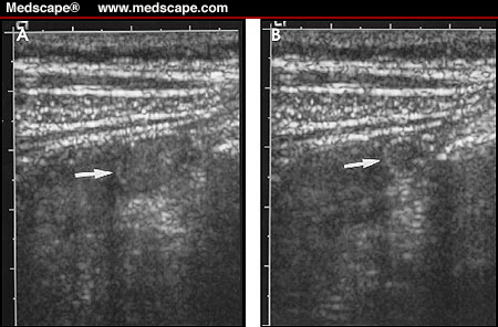
figure 7 Normal bowel. (A) Ultrasound examination of the RLQ demonstrating 1-cm loop of fluid-filled bowel (arrow). (B) With compression and normal peristalsis, the diameter of this normal loop of bowel decreases to 5 mm (arrow).

Ultrasound findings of acute appendicitis are listed in Table 2 . These include a blind-ended, noncompressible fluid-filled structure with a wall thickness of ≥ 3 mm and an outer diameter of ≥ 7 mm identified around the appendix (figure 8). There may be a circumferential color-flow identified around the appendix (figure 9). There may also be free fluid noted in the RLQ of the abdomen and the pelvis, and there may be echogenic mesenteric fat (figure 10). If appendiceal perforation has occurred, an ill-defined and/or fluid-filled abscess may be identified. While 6 mm is usually identified as the cutoff between normal and abnormal appendix, in some instances, 5 mm is used as the upper limits of normal, while 7 mm is considered to be positive for acute appendicitis. Thus, in some instances, a measurement between 5 and 7 mm is considered to be equivocal.
figure 8 Acute appendicitis. Ultrasound of the RLQ of the abdomen demonstrating blind-ended tubular structure (open arrows) corresponding to acutely inflamed appendix. Note the distended lumen [L], the echogenic surrounding mesentery [M], and the echogenic structure with acoustic shadow (arrow) at the base of the appendix corresponding to an appendolith.


figure9 Acute appendicitis. Transverse color-flow ultrasound of the RLQ of the abdomen demonstrates increased vascularity (arrow) in a fluid-filled structure corre-sponding to acute appendicitis. External iliac artery [A] is identified.


figure 10 Acute appendicitis. Ultrasound demon-strating a blind-ended tubular structure with fluid-filled lumen [L]. Also note there is a small amount of fluid [M] surrounding the tip of the appendix.


http://www.medscape.com/viewarticle/431119_4


Appendiceal abscess drainage. A. A 19-year-old who presented with fevers and right lower quadrant pain. CT scan shows an appendiceal abscess (arrows). B. Following ultrasound guided needle placement, a guidewire is coiled in the cavity under fluoroscopy. C. An 8-Fr drain is placed under fluoroscopic guidance and the abscess is drained. D. Follow-up CT scan after catheter removal shows complete resolution of the abscess.
http://www.radrounds.com/.../listTagged?tag=abscess
http:// imaging.consult.com/imageSearch?query=sagitta...
imaging.consult.com/imageSearch?query=sagitta...

Sunday, June 27, 2010

1) A line ① is drawn along the top of the vertebral body of the lower spinal segment.
2) Then at the top-back most portion of the lower vertebral body, draw line ② at 90 degrees to line ①, till it projects well into the body of the vertebra above.
3) Then draw another line ③ parallel to the line just drawn ② this time at the posterior most lower portion of the upper vertebral body.
4) The distance between the upright lines ② and ③ is measured. Any distance of 2mm or greater is a retrolisthesis. This measurement represents the degree of translation (slippage) of the upper of the two segments. See Photo.

http:// headbacktohealth.com/Retrolisthesis.html
To determine the grade of spondylolisthesis, or slippage, the disc is divided in quarters. The grade is equal to the number of quarters of slippage. If there is no slippage, the grade is zero. If the slippage is equal to one quarter of the total width of the disc, the slippage is grade one. If the slippage is three quarters of the width of a disc, it is a grade three spondylolisthesis. If the slippage is more than four quarters (the whole disc space) then it is called a grade 5. In a grade 5 spondylolisthesis, the spine is completely dislocated.

http://www.neurosurgical.com/.../spondylolysis.htm

cobb's angle


which is a sagittal (from the side view) T2 Weighted MRI lumbar image, demonstrates two types of disc herniation: the L5/S1 disc has suffered a 9mm disc extrusion (red arrow) that is not contained by the PLL. The L4/5 disc has suffered a smaller 4mm disc protrusion (green arrow) that is contained by the PLL. The L3/4 (blue arrow) is completely normal and has no disc material projecting posteriorly into the epidural space. Also note that the L3/4 disc is white in color, which indicates it is non-degenerated (i.e., full of water and healthy proteoglycan). The two herniated discs (L4/5 & L5/S1) are "black" on this MRI image, which indicates disc desiccation (lack of water and proteoglycan) and is termed "degenerative disc disease" (DDD), which is usually a precursor to disc herniation for it weakens the annulus which contains the pressurized nuclear material.

http://www.blogger.com/www.chirogeek.com/001_tutorial_birth_of_hnp.htm

Tuesday, June 22, 2010


1) hypoechoic thyroid gland 2) coarse echotexture of the gland 3) fine linear echoes within the thyroid parenchyma s/o fibrosis 4) Color doppler imaging reveals augmentation of the vascularity of the thyroid gland. These ultrasound images are diagnostic of Hashimoto's thyroiditis.
thyroiditis:enlarged thyroid gland(arrows) with diffusely coarsed hypo echoic echotcture
that is typical of Hashimoto's thyroiditis.


scanty blood flow seen in this normal thyroid scan.
marked hyper vascularity of the left thyroid parenchyma in case of grave's disease.

On ultrasound examination, the enlarged and asymmetric gland usually contains multiple solid nodules 1 to 4 cm large. Coarse calcifications may be present, as may areas of hemorrhage or necrosis. A solitary nodule found on clinical or scintigraphic examination has been considered more likely to be neoplastic than multiple nodules.5 On pathologic examination, however, half of clinically identified single nodules have proved to be dominant nodules of multinodular goiters.22 In another study,23 33% of patients undergoing surgical treatment for papillary cancer had coexistent benign nodules found intraoperatively. Therefore, multinodularity does not mean benign disease. Every nodule must be examined for its own ultrasonographic characteristics. As with the physical examination, if ultrasonography can identify a single dominant or more malignant-appearing nodule in a multinodular gland, FNA is directed to the more suspicious-looking nodule. A palpable or dominant nodule should particularly be suspect.
two hypo echoic nodules in the right and left thyroid lobes,the one seen in the right lobe showed punctate psammomatous calcification(small arrows).
benign adenoma showed large amount of blood flow.
spectral doppler showed low peak systolic velocity and prominent diastolic flow.
papillary carcinoma showed moderate amount of blood flow.
spectral doppler showed higher peak systolic velocity with relatively less diastolic flow than within benign nodule.

thyroid nodule with calcified rim(the presence of internal coarse or peripheral eggshell-type calcifications suggest benign lesion ).

Monday, June 21, 2010


1cm hyper echogenic nodule


Other benign features include hyperechogenicity relative to the adjacent parenchyma
Benign cystic nodule with dependent debris.

Typical benign nodules are well-marginated, mostly cystic masses that almost always contain internal debris
In multiloculated benign cystic lesions, internal septa may be thickened but should be devoid of vascular flow.