Best Bath Store
All Natural Blend of 14 Herbs Gentle Cleanse- $20 Off Colonix Now! Use Promo Code 20J
Amberen - Breakthrough Menopause Treatment
728x90
Custom Search

Saturday, July 17, 2010


Schematic representation of the topography of the main groups of perforating veins (PVs). Foot PVs: 1.1, dorsal foot PV; 1.2, medial foot PV; 1.3, lateral foot PV. Ankle PVs: 2.1, medial ankle PV; 2.2, anterior ankle PV; 2.3, lateral ankle PV. Leg PVs: 3.1.1, paratibial PV; 3.1.2, posterior tibial PV; 3.2, anterior leg PV; 3.3, lateral leg PV; 3.4.1, medial gastrocnemius PV; 3.4.2, lateral gastrocnemius PV; 3.4.3, intergemellar PV; 3.4.4, para-achillean PV. Knee PVs: 4.1, medial knee PV; 4.2, suprapatellar PV; 4.3, lateral knee PV; 4.4, infrapatellar PV; 4.5, popliteal fossa PV. Thigh PVs: 5.1.1, PV of the femoral canal; 5.1.2, inguinal PV; 5.2, anterior thigh PV; 5.3, lateral thigh PV; 5.4.1, posteromedial thigh PV; 5.4.2, sciatic PV; 5.4.3, posterolateral thigh PV; 5.5, pudendal PV. Gluteal PVs: 6.1, superior gluteal PV; 6.2, midgluteal PV; 6.3, lower gluteal PV.

Sensitivity and Specificity of Colour Duplex Ultrasonography


The accuracy of colour duplex USG in detecting SFJ and SPJ incompetence was 100%, while 92 perforators could be detected out of 104 by this modality (88.4% sensitivity) and 12 sites were missed, which were situated mainly in the lower part of the leg. By colour duplex sonography, a total of 100 sites were marked as incompetent perforators, out of which 92 were found correct on exploration (92% specificity). So, for perforator incompetence, colour duplex USG missed detection of 12 perforator sites, and 8 sites were marked wrongly as false positive (Table II).
Some perforating vein groups important for surgical treatment of venous insufficiency, seen from behind. (Dodd's group are venae comm. fem. med. intermedia, Boyd's are the vv. comm. cruris intermed., and Cockett's group empty into a vein of the vv. tib. post.). The levels of two dorsal perforators are also indicated, the upper communicating with a gastrocnemius vein.
Schematic drawing of the left pelvic and lower extremity veins. The deep leg veins from the level of the popliteal vein are paired (not shown). The gastrocnemius veins are paired and duplicated; there are several soleal veins.


www.medcyclopaedia.com/.../nic_k201_000.jpg

surgical anatomy of saphenous veins

Figure 1. Superficial veins: LSV; long saphenous vein, SSV; short saphenous vein, SFJ; saphenofemoral junction, PAV; posterior arch vein, SPJ; saphenopopliteal junction. (Picture drawn by Pentti Rautio)
Figure 2. Anatomy of the right saphenofemoral junction:
AL; anterolateral tributary,
FV; femoral vein,
IL; inguinal ligament,
PM; posteromedial tributary,
SCI; superficial circumflex iliac vein,
SE; inferior superficial epigastric vein,
SEP; superficial external pudendal vein. (Picture drawn by Pentti Rautio)

occluded great saphenous vein

Sonogram and cartoon showing an open saphenofemoral junction with a short patent segment above an otherwise occluded great saphenous vein (GSV). The superficial external pudendal (SEP) vein is patent with normal, prograde flow through the SFJ. CFV, Common femoral vein.


http://www.google.com.eg/imgres?imgurl=http://www.veinsurg.com/gal/biblio/chir/chir_7/fig1.jpg&imgrefurl=http://www.veinsurg.com/fr/biblio/chirurgie/chirurgie_7.php&usg=__kayhyYvmS0h6O5tMoixfyZxDgCg=&h=561&w=808&sz=119&hl=ar&start=20&tbnid=wQdy4jSjGeefoM:&tbnh=99&tbnw=143&prev=/images%3Fq%3Dhunterian%2Bperforator%26hl%3Dar%26sa%3DG%26gbv%3D2%26tbs%3Disch:1&itbs=1

great saphenous vein reflux

great saphenous vein reflux
a, Pretreatment duplex image showing midthigh great saphenous vein reflux. Note diameter markers. b, Same vein level at 6 mo, showing diameter reduction, vein wall thickening, and a narrow, irregular, echolucent lumen without flow. c, Same level at 2 years, now seen as a featureless hyperechogenic stripe with further diameter shrinkage and no discernible lumen.


http://www.google.com.eg/imgres?imgurl=http://www.veinsurg.com/gal/biblio/chir/chir_7/fig1.jpg&imgrefurl=http://www.veinsurg.com/fr/biblio/chirurgie/chirurgie_7.php&usg=__kayhyYvmS0h6O5tMoixfyZxDgCg=&h=561&w=808&sz=119&hl=ar&start=20&tbnid=wQdy4jSjGeefoM:&tbnh=99&tbnw=143&prev=/images%3Fq%3Dhunterian%2Bperforator%26hl%3Dar%26sa%3DG%26gbv%3D2%26tbs%3Disch:1&itbs=1
Small Saphenous Vein (SSV)
Courses from the lateral ankle up the posterior calf
Terminates in the popliteal fossa at the saphenopopliteal junction (SPJ)
Confluence with the popliteal vein (PV) is variable
Proximal portion lies between superficial & deep fascial layers


http://www.google.com.eg/imgres?imgurl=http://www.icvein.com/images/saphenous-vein.jpg&imgrefurl=http://www.icvein.com/venous-disease-varicose-vein-treatment-iowa-city-ia.htm&usg=__Ne7smwjRmou-16vDYSuoafZgyNI=&h=385&w=221&sz=19&hl=ar&start=3&tbnid=jKodCR2axztKAM:&tbnh=123&tbnw=71&prev=/images%3Fq%3Dlesser%2Bsaphenous%2Bvein%2Bperforators%26hl%3Dar%26sa%3DG%26gbv%3D2%26tbs%3Disch:1&itbs=1
Connect superficial to deep veins
Locations
Proximal thigh - Hunterian
Distal thigh - Dodd’s
Knee - Boyd’s
Ankle/Calf - Cockett’s
Incompetent perforators often source of venous stasis ulcers at medial ankle


http://www.google.com.eg/imgres?imgurl=http://www.icvein.com/images/saphenous-vein.jpg&imgrefurl=http://www.icvein.com/venous-disease-varicose-vein-treatment-iowa-city-ia.htm&usg=__Ne7smwjRmou-16vDYSuoafZgyNI=&h=385&w=221&sz=19&hl=ar&start=3&tbnid=jKodCR2axztKAM:&tbnh=123&tbnw=71&prev=/images%3Fq%3Dlesser%2Bsaphenous%2Bvein%2Bperforators%26hl%3Dar%26sa%3DG%26gbv%3D2%26tbs%3Disch:1&itbs=1
emedicine.medscape.com/article/462579-overview‎

The venous network in the lower extremities commonly affected by CVI is divided into 3 systems. The first is superficial veins, which include the lesser and greater saphenous veins and their tributaries, as depicted in the 1st image below. The second is deep veins, which include the anterior tibial, posterior tibial, peroneal, popliteal, deep femoral, superficial femoral, and iliac veins. The third is perforating or communicating veins, as depicted in the 2nd and 3rd image below.


In patients with symptomatic greater saphenous varicosities, the presence of an occluded deep system must be ruled out. Deep occlusion is an absolute contraindication to vein ligation. Obtaining venographic studies of the deep venous system prior to superficial vein ligation is imperative.

1-Anatomy
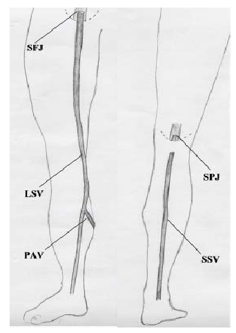
The superficial veins lie in the subcutaneous fatty layer of the body just beneath the skin and superficial to the deep fascia enveloping the body musculature. The principal veins in the legs are the great and lesser saphenous veins and their tributaries; in the arms they are the basilic and cephalic veins and their tributaries. The deep veins accompany arteries and bear the same name as the arteries they parallel. It is common in the extremities for there to be two or more veins accompanying a small- to medium-sized artery. The perforating veins penetrate the deep fascia and connect the superficial veins to the deep veins. Those along the inner (medial) side of the lower leg play a major role in the pathogenesis of the "postphlebitic leg". The intra-muscular sinusoidal veins are large, very thin walled, valveless veins within skeletal muscle. They connect directly with the deep veins.
2-Physiology
The venous system of vessels conducts blood back to the heart. Normal venous flow is dependent on four factors: the heart (dynamics/spontaneous flow), respiration (phasic flow), the venous pump and the valves.
Dynamic Flow: Flow in the arterial system is dependent on the pumping action of the heart and the elasticity and muscular activity of the arteries. In contrast, the veins, except for the major superficial veins, are thin walled with paucity of muscle; are designed for distention; and play an insignificant role in directly facilitating flow. Most of the force of the pulsatile flow produced by the pumping heart is lost as blood flows from the arteries through the vast network of capillaries (one cell layer micro-vessels where selective permeability allows the exchange of O2 and nutrients with the tissues). What pulsatile flow filters through (dynamic flow) is of low pressure (15mmHg). Nevertheless, it is sufficient to produce a significant pressure gradient with the right side of the heart where the venous pressure is 0.
Phasic Flow: is the effect of respiration on normal venous flow. In the arms and neck, flow towards the heart increases during inspiration due to the negative intra-thoracic pressure produced. The opposite is true in the legs. With inspiration the diaphragm descends increasing intra-abdominal pressure slowing flow. Flow is somewhat increased with expiration with reduced intra-abdominal pressure.
The "Muscle Pump": the muscle pump mechanism is most highly developed in the calf muscles. Large venous sinusoids located in these muscles act as a bellows and the contracting muscle the force emptying the bellows. Contractions of the calf muscles can produce a pressure in excess of 200 mmHg. This is sufficient pressure to empty the blood out of the sinusoids into the deep veins. The deep veins in turn are subject to a similar compressing force because of the strong fascial investment about the muscle compartment in which they are contained. As a result blood is pumped towards the heart with each muscle contraction (diagram #3).
Valves: valves are structured so that flow is always towards the heart and flow from superficial to deep veins. Without valves there would be one continuous column of blood from heart to ankle when an individual stood. By preventing reflux the valves complement the muscle pump in returning blood to the heart.

3-pathology
The most common pathological conditions in the venous system occur in the legs, and include valve incompetence and venous obstruction.
Venous obstruction is most commonly due to venous thrombosis, but may also result from vein compression (tumors, cervical rib, fractures, hematomas, arterial aneurysms etc). Factors producing venous thrombosis are vein injury, flow stasis (inactive muscle pump from lack of motion, bed confinement or paralysis), blood hypercoagulability (in post operative patient or an intrinsic coagulation disorder) and a combination of above as may occur in multiple trauma patients.
Valve incompetence may be due to congenital valve defect or develop as a complication of venous thrombosis and venous hypertension. Congenital valve incompetence occurs in the superficial veins and the perforators. Deep vein valve incompetence is considered a complication of previous deep vein thrombosis regardless that a history of deep vein thrombosis is obtained in less than 50% of patients.
Incompetence results in reflux with increased venous pressure in the segment of vein(s) below the incompetent valves when the individual stands (a condition referred to as "venous insufficiency"). Clinical manifestations depend on which venous system(s) (superficial, deep or perforators) are involved and the number of valves incompetent. When the valves of the deep veins are affected the ensuing venous hypertension produces backpressure in the capillaries causing leakage of fluid into the tissues with leg edema (swelling).
4-Venous Diseases
a-Varicose Veins
A varicose vein is dilated, elongated and tortuous. Most commonly affected are the saphenous veins in the legs (90% involve great saphenous system). The cause is venous hypertension resulting from valve incompetence. They may be primary or secondary.
Primary (saphenous) varicose veins
This is a congenital condition. Varices result from congenital weakness of the valve structure and possibly also a congenital weakness in the vein wall. There is a family history in 75% of patients. By themselves primary varicose veins produce few symptoms. Cosmetic appearance is the major complaint. A common symptom is heaviness of the legs towards the end of the day, particularly for those whose occupation requires considerable walking and standing. There should be little to no ankle or leg swelling.
Secondary Varicose Veins
Secondary varices are a sequela of either deep vein obstruction, incompetent deep vein valves or a combination of both. In each case the resulting venous hypertension renders the perforating veins incompetent allowing unrestricted back flow from deep to superficial veins. The superficial veins not being structured to withstand a high venous pressure become dilated and elongated, forming secondary varices.
Thrombophlebitis, Venous Thrombosis
When a thrombus (blood clot) obstructs a vein it sets up a sterile inflammatory reaction in the vein wall and the surrounding tissue. This condition is known as "thrombophlebitis". When a loosely attached thrombus develops in a vein and is not obstructing, it produces no reaction in the vein wall. This condition is known as "phlebothrombosis."
Superficial Thrombophlebitis:
Superficial thrombophlebitis of the great saphenous vein presents with a typical clinical picture. It begins with sudden development of pain and tenderness along the course of the section of vein involved. The skin over the vein is red and the adjacent tissue swollen. With resolution of the inflammation (2-3 weeks if untreated) the thrombosed vein can be felt as a cord-like structure beneath the skin.
The cause of the phlebitis and the reason that the great saphenous vein is usually targeted remains unknown. Local trauma and/or unusual activity may be a precipitating cause in some cases. It can be recurrent, developing in another section of the saphenous vein in the same leg. Superficial phlebitis is more of an annoyance than a serious condition. It resolves rapidly with reduced activity and anti-inflammatory drugs. However, in those cases where the thrombotic process extends to the level of the groin, there is a risk of involvement of the common femoral vein and the possibility of pulmonary embolism.
Superficial saphenous vein thrombophlebitis is distinct from thrombosis occurring in a cluster of varices. The latter is common in varicose vein disease. There is no risk of pulmonary embolism. Permanent discolouration of the overlying skin is the main complication. In the arms the common cause of superficial phlebitis is chemical damage to the lining of the vein from intravenous injection. Pulmonary embolism secondary to thrombophlebitis in the arms is rare.
Deep venous thrombosis (D.V.T)
D.V.T. by medical convention refers to thrombosis in the deep veins of the legs. It is a relatively common complication of major surgery, leg fractures and prolonged bed rest. Stasis because of muscle pump inactivity, a hypercoagulable state which is a biological reaction to injury and, local trauma to veins, all play a role.
The clinical presentation is related to site, extent and degree of obstruction produced by the thrombus. Thrombosis limited to the calf veins produces only mild calf soreness and tenderness and minimal, if any, ankle swelling. In contrast acute thrombosis obstructing the femoral and iliac veins results in a grossly swollen, painful, white leg (white because of the extensive edema under the skin). Rarely the extent of the thrombosis will include the entire venous network causing obstruction of arterial circulation. The result is massive leg swelling complicated by manifestations of vascular ischemia and possible gangrene (so called venous gangrene). When deep vein thrombosis does not cause obstruction (i.e. phlebothrombosis) pulmonary embolism may be the first and only clinical manifestation.
Acute D.V.T. is a medical emergency. Anticoagulation (blood thinning) is the prime treatment. It prevents extension of the thrombus. Clot lysing drugs (thrombokinins) are injected transvenously in selected cases. Thrombectomy (surgical removal of clot) has a limited role.
Recurrence of acute D.V.T. is common. Major complications include pulmonary embolism, chronic deep vein insufficiency and the postphlebitic leg.
Pulmonary Embolism
Pulmonary embolism occurs when a piece of clot (thrombus) breaks away from the vein wall, enters the venous flow and passes through the right side of the heart to lodge in the pulmonary artery or one of its branches. Clinical manifestations are determined by the size of the clot and the size of the vessel occluded. The spectrum includes sudden death (occlusion of main pulmonary artery), pleurisy-like symptoms from lung infarction (occlusion of segmental artery and local lung death); shortness of breath and pulmonary insufficiency (occlusion of multiple small vessels from showers of small emboli). Acute D.V.T. is the major source of pulmonary embolism.
Lung scans and pulmonary angiograms confirm the diagnosis. Immediate and full anticoagulation (blood thinning) is basic treatment. Where obstruction of the main pulmonary or major branch is diagnosed thrombolytic agents are injected directly into the clot by intravenous catheter. Surgical removal of the blood clots is warranted in selected cases. When the use of anticoagulants, is contraindicated a filter is inserted into the inferior vena cava to prevent further clots from reaching the lung.
Chronic Venous Insufficiency and Postphlebtic Leg
Chronic venous insufficiency is an overarching term which includes the various clinical and pathological entities resulting from impaired venous flow and venous hypertension. Chronic venous insufficiency in the deep veins is attributable to acute D.V.T. and its sequelae. The degree of insufficiency initially depends on the fate of the obstructing thrombus. The thrombus may be partially or completely dissolved by lytic enzymes. More commonly it is organized and replaced by fibrous tissue with varying degrees of recanalization (the development of small, valveless channels traversing the length of the organized thrombus). Recanalization produces little improvement in venous flow. Compensatory dilatation of collateral veins (superficial and deep secondary veins which bypass the obstructed area) is more effective in re-establishing the venous flow. The dilated superficial veins which may result are referred to as secondary varicose veins.
D.V.T. has a disastrous effect on the valves of the deep and perforating veins partially due to injury to the valves by the process of thrombosis, and more particularly due to venous hypertension, which produces abnormal dilatation of both the deep and perforating veins rendering their valves incompetent. The result is the inability of the venous pump to provide relief from venous hypertension when the individual stands and walks. As a consequence, the capillaries in the distal part of the legs are exposed to a high pressure (Diagram #4).
The result is increased capillary permeability with leakage of fluids into the tissues producing leg edema (swelling). The concentration of perforators around the ankle and the higher hydrostatic pressure with standing makes this area (the so-called "gaiter area") particularly vulnerable. Diagram 5


Pathophysiology

To ensure adequate venous return from the lower limbs, the superficial veins, deep veins, bicuspid valves and the calf muscle must all work together [7]. The deep veins carry blood back up the leg and consist of two posterior tibial veins, two anterior tibial veins and two peroneal veins which join up to form the popliteal vein which in turn continues into the femoral veins. These veins are situated deep in the muscles of the leg and are protected by a fibrous fascia. The large volume of blood carried by these veins results in a high pressure within the vein walls [8] whereas the superficial veins, consisting of the long saphenous veins, short saphenous veins and numerous superficial collaterals, satellites and confluents of the saphenous veins (Figure 1), carry a smaller volume of blood at a lower pressure. The superficial veins drain into the deep veins by means of the perforating veins. Bicuspid valves, present in both superficial veins and deep veins, ensure that the flow of blood is unidirectional and when these valves are competent they prevent a backflow of blood from the deep veins to the superficial veins.

Figure 1 - Long and short saphenous system

The power to drive the blood back up the leg is provided by the calf muscle, which on walking contracts and relaxes in a regular movement. The contraction of the calf muscle forces the blood upward out of a segment of vein; backflow is prevented by the valve [7]. Relaxation of the calf muscle allows the now empty segment of deep vein to refill with blood from the superficial veins and thus the cycle is repeated.

When valves become incompetent the cycle of unidirectional blood flow is interrupted and backflow of blood occurs (Figure 2). This is most significant when the backflow occurs between the deep and superficial veins, as the increased pressure in the superficial veins will cause further valve incompetence. This is because the valve cusps no longer meet as a result of the stretching of the veins. The overall effect of this increased superficial hydrostatic pressure is the formation of tortuous varicose veins [9].

Figure 2 - Venous system (normal and damaged)


www.worldwidewounds.com/2002/september/Johnso...‎

Friday, July 16, 2010

Measuring the ankle-brachial index (ABI). The ABI is 95% sensitive and 99% specific for angiographically measured lower extremity arterial stenosis of 50% or greater. The ABI is calculated as the ratio of Doppler recorded systolic pressures in the lower and upper extremities. DP=dorsalis pedis; PT=posterior tibial. Adapted with permission of the Massachusetts Medical Society from N Engl J Med. 2001;344:1608-1621.[47]

the ankle-brachial index (ABI) is highly sensitive and specific for angiographically confirmed PAD.[22-25] The ABI is a ratio of Doppler-recorded systolic pressures in the lower and upper extremities. The ABI requires approximately 10 minutes to perform. It is a noninvasive and reliable measure of the presence and severity of PAD. To measure the ABI, systolic pressures are measured in the brachial arteries of the upper extremities and in the dorsalis pedis and posterior tibial arteries of each lower extremity with a hand-held Doppler (Figure 1). The ABI is calculated for each lower extremity by dividing the average of the posterior tibial and dorsalis pedis arterial pressures in each leg by the average of the right and left brachial artery pressures.[26] In a person without PAD, arterial pressures increase with increasing distance from the heart, resulting in an ABI >1.0.
site of examination of the dorsalis pedis.
site of examination of the posterior tibial artery.

how is the spectral wave form appeared.

Assessing the lower-limb arteries

Blood is normally supplied to the leg through a single main artery (Figure 7). This has different names in different parts of the leg: common and external iliac arteries in the lower abdomen, common and superficial femoral arteries in the thigh, and popliteal artery behind the knee. Below the knee it branches into the smaller posterior tibial, peroneal and anterior tibial arteries. The reason for scanning these arteries is to locate and assess any narrowing (stenosis) or blockage (occlusion). A general review of the role of ultrasonic scanning in the diagnosis of peripheral arterial disease is given by Polak [4] .

Figure 7: The main arteries of the lower limb.

Patients are normally referred with claudication (pain on walking, particularly uphill) or with ulcers around the ankle and on clinical examination the ankle pulses are likely to be weak or absent. The main options for treatment are either (a) to encourage the patient to exercise and develop collateral vessels to take blood around the site of disease, (b) to perform angioplasty which involves passing a balloon catheter down the artery and inflating it at the sites of narrowing or blockage to dilate the vessel, or (c) to surgically insert an graft to bypass the diseased portion of the vessel [5] . The clinical information required is therefore the site and severity of any narrowing of the vessel and the site and length of any blockages of the main vessels. In the presence of multiple stenoses, the study can indicate which are causing the more critical restrictions to blood flow. The referring clinician can use this information to decide whether intervention is appropriate, and, if so, whether to proceed to angioplasty or to insert a graft [6] .

The examination starts with the patient lying supine with the head slightly raised. Coupling gel is put on the thigh from groin to knee over the path of the artery. The probe is placed on the skin in the groin and the common femoral artery identified using the colour Doppler display. The scanner is switched to duplex mode, and the blood velocity waveform in the common femoral artery is obtained. The waveform is usually biphasic or triphasic. The peak systolic blood velocity normally lies between 90 and 140 cm/s (ref 3, p 260). Values significantly above this may indicate local stenosis, while values below can indicate low flow caused by proximal or distal occlusion. The presence of any plaque intruding into the lumen is noted, and the degree of stenosis is estimated. (Figure 8) shows a scan of a femoral artery with a small protruding plaque causing about 20% stenosis.


Figure 8: A plaque protruding into the lumen of the common femoral artery. The plaque (PL) is small and is causing about 20% stenosis.

The shape of the waveform is important because a monophasic waveform can indicate proximal disease in the iliac vessels. The biphasic or triphasic waveform occurs because the main blood vessels in the leg are elastic and are dilated by the increased pressure during systole. This creates a reservoir of blood which empties during diastole. The volume of blood in the reservoir is more than enough to supply the limb, and the excess flows back up the vessel into the abdominal aorta creating the reverse flow component (Figure 9). An abnormal monophasic waveform without the reverse component occurs when the volume in the reservoir is insufficient and extra flow is required during diastole. This is usually because an iliac stenosis or occlusion reduces the blood available to fill the reservoir during systole, but may also occur when there is a large flow to the limb caused by exercise or gross infection.

Proximal disease can also be indicated by turbulence in the common femoral artery. Turbulence is created in eddies distal to a tight stenosis, and the eddies then travel downstream with the blood flow. They can be detected as spikes in the blood velocity waveform (Figure 10), usually on the downslope at the end of systole. The turbulence is more likely to be created during peak systole when the local blood velocity is highest, but appears later than this distally because the systolic pressure pulse travels faster than the eddying blood. The delay between peak systole and the appearance of the turbulence in the common femoral artery can give an indication of the site of the proximal iliac stenosis. Jager et al [7] have described the shape of the waveform in the lower-limb arteries and the changes associated with different degrees of stenosis.


Figure 9: Schematic generation of a biphasic waveform. During systole (a), the heart pumps blood into the peripheral vessel and the branches. The peripheral vessel expands. During diastole (b), the vessel contracts. There is more than enough blood in the vessel to supply the periphery, and the excess flows backwards into the more proximal branches. A biphasic waveform is obtained at the point marked with an asterisk.


Figure 10: Turbulence in the common femoral artery. The blood velocity waveform contains spikes (marked I) just after peak systole. These represent turbulence generated from a proximal tight stenosis
The probe is then moved along the artery until the origin of the profunda femoris artery is identified using the colour Doppler display. This is usually just beyond the skin crease in the groin. The blood velocity waveform at the origin is obtained, the waveform shape noted and the presence of any stenosis recorded. The position of the origin can be marked on the skin surface using a water-soluble crayon.
The superficial femoral artery is examined along its length using the colour Doppler display. The colour scale is set so that the normal blood velocity in the vessel is just below the top of the scale. Any increase in velocity caused by a stenosis will therefore go above the scale, causing aliasing and its characteristic display. Whenever a stenosis is suspected, the blood velocity waveform is obtained just proximally and the peak systolic velocity measured. The duplex gate is then moved through the stenosis, monitoring the waveform until the maximum velocity is obtained. A significant stenosis is normally taken to be one that more than doubles the blood velocity [4]. The site of any significant stenosis is marked on the skin surface and the distance from the vessel origin and the increase in velocity recorded. Figure 11 shows a superficial femoral artery stenosis. The plaque causing the stenosis can be seen and the colour Doppler shows aliasing. The peak systolic blood velocity, (Figure 12), increases from 0.26 proximally to 3.90 m/s through the stenosis, an increase by a factor of 15 indicating a very tight stenosis.
Occlusion is characterized by a gradual fall in blood velocity along the vessel as blood is taken away by collateral vessels. The waveform usually becomes monophasic close to the occlusion, and at the blockage the colour Doppler display shows absence of flow. When flow is low, it can be difficult to determine the precise point at which flow ceases, and the operator must ensure that any flow shown is within the vessel rather than along a nearby collateral. When an occlusion is detected, the operator marks the start on the skin, and then continues to track down the artery using the colour Doppler display until flow is again identified in the lumen. The distal end of the occlusion is marked and the length measured.
As the scan progresses down the leg, the artery becomes deeper and can be difficult to image, particularly in larger patients. The vessel is scanned as far down the leg as possible and the blood velocity waveform recorded from this distal site. The position is marked on the skin. To complete the examination of the lower-limb arteries, the patient turns onto their side or front and the popliteal artery is scanned behind the knee. Waveform shape, peak systolic velocity and the presence of plaque are all recorded.
Where the distal superficial femoral artery is deep, there may be a portion of the vessel that is not accessible from the front or the back. This can limit the accuracy of the scan, especially in the adductor canal just above the knee. Problems can also be caused by a vessel with multiple calcified plaques, which block the ultrasound preventing a clear view of the vessel. However, an experienced operator can use other clues such as the presence of turbulence and changes in waveform shape to determine the presence of significant disease. Where there is an occlusion or tight stenosis, collateral vessels re-entering distally can supply blood at high velocity which can mimic a stenosis. The collateral vessel can sometimes be identified by the presence of colour outside the main lumen.


Figure 11: A superficial femoral artery stenosis. The colour image shows aliasing, a sharp transition from maximum speed away from the probe (light blue) to maximum towards (yellow). The lumen is narrower at the stenosis and the brighter echoes in the near wall show the presence of plaque, which is causing shadowing to the right of the image.


Figure 12: Velocity waveform through a stenosis. The peak blood velocity through this superficial femoral artery stenosis is greater than 3.9 m/s, compared with 0.26 m/s just proximal to the stenosis. This increase in velocity shows a very tight stenosis.

A full lower-limb scan may be time-consuming since the whole length of the artery must be scanned carefully. About 30 minutes is needed for a full unilateral study and 50 for a bilateral study. Other tests are sometimes performed at the same time, and it is common to include a measurement of the ankle/brachial pressure index (ABPI) which extends the study by about 10 minutes [8] [9] [10] .
The ABPI (ratio of ankle arterial systolic blood pressure to brachial pressure) is normally greater than 1.0, and ratios lower than 0.90 at rest are usually considered abnormal and indicative of lower-limb arterial disease [8]. However, an artifactually low reading may be obtained if the ankle signals are weak and of low amplitude. Headphones can be useful to ensure that the signal is heard as soon as it returns as the cuff pressure is reduced. Artifactually high readings may also be obtained if the leg arteries are calcified and cannot be compressed. This happens particularly in diabetic patients. In these cases, the arterial waveform at the ankle can help to exclude disease since a biphasic waveform, with both forward and reverse components, indicates absence of severe flow-limiting disease. For all these pressure measurements, it is important that the cuff is the correct size [11] . Ideally, the ratio of cuff width to limb circumference should be around 0.4, and it is good practice to have a large cuff available for large or swollen limbs. Reducing errors is especially important if ABPI is the only measurement being made and the patient is not proceeding to duplex scan.
Proximal and distal arterial scanning

If iliac disease is suspected, either from clinical examination or from a monophasic common femoral waveform, the aorto-iliac segment can be scanned [12] . A 3.5 MHz probe is usually used to give better penetration and a wider field of view. Access to the vessels can be difficult if overlying bowel and bowel gas block the ultrasound. Some centres give an enema before scanning this region, but in thin patients a satisfactory examination is often possible without bowel preparation. The operator is again looking for narrowing or blockage of the artery and the same criteria are used. If distal disease is suspected, some of the small arteries below the knee can be examined. The posterior tibial artery is usually accessible just above the ankle from the medial aspect, and can be traced up the leg as it gets deeper. The artery runs parallel to a pair of veins (Figure 13) and this can aid identification. The operator is again looking for evidence of stenosis or occlusion, but these vessels are small and close to the limit of resolution of all but the most modern scanners, and absence of detected flow may indicate lack of sensitivity of the equipment rather than occlusion.

Figure 13: Peroneal artery and paired veins. The arterial flow, shown in red, is from left to right with a component towards the probe. The venous flow, shown in blue, is in the opposite direction. The anterior and posterior tibial arteries also lie between paired veins.


http://www.blogger.com/www.worldwidewounds.com/.../Doppler-Imaging.html

Assessing the lower-limb veins for incompetence

The lower-limb venous anatomy is complex and variable, but the veins can be divided into deep veins, superficial veins and perforators. The purpose of the veins is to return blood back to the heart, and when standing this has to be done against gravity. To assist the venous return, there is a pump operated by the calf muscles. As these contract, they squeeze blood out of the calf and up the leg. One-way valves in the veins prevent the blood from falling back as the muscles relax. The muscle pump is more effective in the deep veins, so the venous anatomy directs the blood from the superficial veins to the deep veins. (Figure 14) shows the main veins in the calf and thigh: the common femoral, superficial femoral, popliteal, posterior and anterior tibial and peroneal veins are all part of the deep system. The main superficial veins are the long and short saphenous veins, and there are also the gastrocnemius and posterior thigh veins. The long and short saphenous veins join the deep veins at the sapheno-femoral and sapheno-popliteal junctions respectively. Perforators link the superficial and deep systems, and contain valves to allow flow only from superficial to deep. Normal venous anatomy is variable and becomes more so in disease when superficial veins enlarge and become varicose. Some of the variations seen on duplex scanning have been described by Somjen


Figure 14: The main veins of the lower limb. The long and short saphenous veins are superficial veins and the remainder are deep veins.


The main reason for scanning these veins is to detect veins in which the valves leak. The leakage may be due to valvular damage or to venous distension [14] . If the valves leak, the veins become incompetent and the blood falls back under gravity as the calf muscles relax, increasing the venous pressure because of the hydrostatic pressure of the column of blood being supported. The condition is known as venous insufficiency. Persistent increased pressure causes superficial veins to dilate producing varicosities, and causing tissue damage distally, showing first as changes in skin colour and progressing to ulceration. The main purpose of treatment is to reduce the excess venous pressure, and this can be done surgically or using bandages. Surgical treatment removes or ties the incompetent veins, and is suitable for superficial vein incompetence since there are other veins which can carry the venous return. The veins are tied at all points where the higher pressure blood from the deep veins enters the superficial system. This is usually at the sapheno-femoral junction or the sapheno-popliteal junction or through incompetent perforators linking the deep and superficial systems. However, the deep veins cannot be removed or tied since they are required to return the blood to the heart. If the deep veins are incompetent, the leg is bandaged to increase the external pressure so that the tissue pressure more closely matches the venous pressure [15] . The applied pressure is graded, being greater at the ankles and decreasing up the leg to encourage the venous return [16] [17] .

The clinical requirement is therefore to identify any superficial incompetent veins and to locate the points where blood is entering from the deep venous system. The presence or absence of deep vein incompetence must also be determined. If there are several incompetent vessels, it can also be useful to indicate which appear to be more significant. A simple examination can be performed using a hand-held continuous wave Doppler unit [18] but this only gives limited information and a full colour duplex scan is preferable. The duplex technique is described in detail by Polak [3]. Patterns of venous reflux have been described by Myers et al [19] and Lees and Lambert [20] , and correlated with clinical symptoms and signs by Labropoulos et al [21] . Further validation of the duplex colour flow examination has been described by Pierik et al [22] .

The examination starts with the patient standing facing the investigator, or lying supine on a couch tilted feet down at least 20� from the horizontal. This is to ensure the veins are filled, and also to ensure that gravity will return blood through any incompetent veins. Coupling gel is put on the probe, which is placed lightly on the skin in the groin over the femoral vein. A light probe pressure is essential, since too great a pressure can narrow or occlude the vein. The femoral vein is identified using the colour Doppler display, and the probe moved along the vein until the site of the sapheno-femoral junction is located (Figure 15). This is near to the point where the femoral vein comes closest to the skin surface. The colour box is placed on the image of the femoral vein just distal to the junction and the thigh is squeezed gently. Flow should be seen in the vein, the colour indicating flow towards the abdomen.The squeeze is then released, and the image inspected for any reverse flow during the release. Any reverse flow persisting for more than one second is normally taken to indicate significant incompetence (ref 3, p 234), although some workers use 0.5 sec [19] or 0.6 sec [20] as the cut-off. The colour box is then placed over the image of the sapheno-femoral junction where the long saphenous vein meets the femoral vein, and the thigh again squeezed and released (Figure 16). Reverse flow persisting for more than 1 sec indicates significant long saphenous vein incompetence.


Figure 15: Longitudinal scan of a sapheno-femoral junction. The superficial long saphenous vein (LSV) joins the deep superficial femoral vein (SFV) to form the deep common femoral vein (CFV)

Figure 16: A normal sapheno-femoral junction on squeeze/release. The blue in the long saphenous vein shows flow towards the heart. The blood velocity waveform shows flow towards the heart as the thigh is squeezed and the flow continues in the same direction as the squeeze is released.

Although this procedure can seem straightforward, there are conditions which make the study difficult. The thigh can be difficult to squeeze, and in this case a co-operative patient can perform a Valsalva manoeuvre. The patient breathes in, closes their mouth and nose or throat, and increases the abdominal pressure by trying to force air out against the obstruction. The increased pressure is transmitted to the veins, and reverse flow is seen in the femoral or long saphenous veins if these are incompetent.

Some patients may have recurrent incompetence which has developed since previous surgery. In these cases the long saphenous vein (LSV) may have been ligated and may not communicate directly with the femoral vein. However, small collateral veins may have opened, linking the femoral vein to the more distal LSV, or there may be incompetent perforators linking in the same way (Figure 17). The next part of the study is therefore to identify the long saphenous vein at mid-thigh level and to assess the degree of any incompetence in the same way as before. The probe is placed over the long saphenous vein on the antero-medial aspect of the thigh, posterior to the path of the superficial femoral artery. The calf is squeezed and released, and the presence and approximate duration of any reverse flow is noted (Figure 18). If an incompetent LSV is demonstrated, the vein should be traced proximally up the thigh to identify the source of the incompetence and in particular to look for the sites of incompetent perforators, which can be marked on the skin surface with a crayon.


Figure 17: A large incompetent upper thigh perforator. The large perforator joins the deep superficial femoral vein (SFV) to the superficial long saphenous vein (LSV). On release of a thigh or calf squeeze, blood would flow from the deep vein through the incompetent perforator into the superficial system.


Figure 18: An incompetent long saphenous vein. There is normal forward flow on squeezing the lower thigh (SQ), but the flow reverses when the squeeze is released (REL). The reverse flow persists for more than two seconds, indicating significant incompetence.

The patient then turns round to face away from the investigator, and relaxes the leg being examined. The probe is placed behind the knee, the popliteal vein and the sapheno-popliteal junction identified and assessed for incompetence by squeezing and releasing the calf. If superficial incompetence is demonstrated, it is important to identify which vein is incompetent. This is usually the short saphenous vein (SSV), but may also be the posterior thigh vein or the gastrocnemius vein.

Having assessed the main deep and superficial veins, it is important to examine any varicose veins to assess how they are being filled. They are usually filled by reverse flow in an incompetent superficial vein (LSV or SSV) but could be filled directly from incompetent perforators. The probe is placed lightly over the varicose vein, which is then traced up the leg using squeeze/release of the calf or thigh to augment flow and aid identification. Reverse flow on release of the squeeze can usually be seen clearly distally, but this may become harder as the probe is moved up the leg. This is because the varicose vein may be fed by small tortuous incompetent veins which pressurize the system but provide restriction to flow. In this case, squeezing the leg distally forces blood up the leg and this dilates the proximal vein providing a reservoir from which the blood falls back on release of the squeeze. However, more proximally the flow reduces because any reservoir is small or non-existent, and it can be impossible to trace the varicose veins satisfactorily. The veins are often tortuous with many branches and it is important to follow the main channel rather than side branches. The only guide is to try to follow the main incompetent vessel.

At this stage of the examination, a clear picture has hopefully emerged, with the incompetent veins identified and a definite source of the incompetence demonstrated. It is very satisfying when this happens, particularly if the picture is complicated. However, there are times when a satisfactory picture does not emerge, and the report has to reflect this.

The final part of the study is to identify any further incompetent perforators, particularly in the calf. It can be difficult to examine the calf with the patient standing on the floor, so some operators stand patients on a stool. Patients do occasionally become faint and very occasionally collapse without warning, so I prefer to examine the calf with the patient sitting up on a couch with the knee bent so that the calf is about midway between horizontal and vertical. The medial, lateral and posterior aspects of the calf are then scanned in longitudinal mode using the colour Doppler display. Incompetent veins can be identified close to the ankle using squeeze/release of the distal calf, and any incompetent veins tracked up to establish any communication with the deep veins. Sites where blood enters the superficial system on release of the squeeze are marked, and the distances from the lateral or medial malleolus are measured and recorded.

Venous examinations, particularly for recurrent incompetence, can be complex and time-consuming. I generally allow 1 hour for a bilateral scan, and 40 minutes for a unilateral study. More time is required if bandages have to be removed before the study. It is important to make notes of the findings at each stage of the study, and a sketch can sometimes be useful as an aide memoire.


http://www.blogger.com/www.worldwidewounds.com/.../Doppler-Imaging.html


Thursday, July 15, 2010

Cornual pregnancy:


Sonography of the uterus shows a gestation sac of 6 weeks 4 days age, in the right cornu of the uterus. 3-D image of the uterus further confirms the findings. These ultrasound images are diagnostic of cornual pregnancy (a type of ectopic pregnancy). Ultrasound images courtesy of Dr. Latha Natrajan, Bangalore, India.

Bicornuate uterus with gestation sac:

Pregnancy in one horn of uterus with two horns (cornu).
1=one horn with decidual reaction.
2=another horn with empty pattern.


http://www.ultrasound-images.com/early-pregnancy.htm

Wednesday, July 14, 2010

How to spot the Normal ductus venosus:

It is often difficult to spot the ductus venosus among the numerous vessels in the fetal abdomen. I have devised an easy way out.
a) First spot the umbilical vein passing through the fetal abdomen.
b) Switch on the color Doppler function to view the flow of the umbilical vein.

c) Reduce or increase the PRF (pulse repetition frequency) function of the color flow until you spot a prominent but short vessel with MARKED ALIASING (ie: turbulent flow producing a multiple shades in the flow image). This is most likely to be the ductus venosus. Note the location of the vessel, just anterior to the fetal aorta.


d) Now switch on the spectral doppler trace of the vessel. This will give a wavy spectral waveform with 3 waves (d): The S wave, the D wave and the A wave. Note the marked diastolic flow in this waveform. This is diagnostic of a normal Ductus venosus. All images by Joe Antony, MD, using a Toshiba Nemio -XG ultrasound system.

Fetal club hand deformity:


This 2nd trimester fetus shows short forearm bone (radius) with radial deviation of the wrist. The image 2 nd is a B-mode ultrasound image showing this deformity of the hand. The other 3 images are 3-D (3 dimensional) ultrasound images showing the same anomaly in this fetus. These ultrasound images suggest fetal radial club hand anomaly. Radial club hand is the commonest fetal club hand anomaly. It is important to look for other fetal malformations as this anomaly is usually not isolated. Ultrasound images courtesy of Dr. Dilraj Gandhi, India.