Best Bath Store
All Natural Blend of 14 Herbs Gentle Cleanse- $20 Off Colonix Now! Use Promo Code 20J
Amberen - Breakthrough Menopause Treatment
728x90
Custom Search
Showing posts with label emergency radiology. Show all posts

Saturday, July 31, 2010

The CT scans demonstrate a fractured hyoid bone, gas bubbles in an abscess producing mass effect, and narrowing of the airway.

This patient received a blow to the neck and noticed increased pain and difficulty swallowing. The hyoid bone is located between the mandible superiorly and the thyroid cartilage inferiorly. It is a “floating bone,” which is supported by strap muscles in the neck. In turn, it supports the root of the tongue, and allows for a wider range of motion, which may create a wider range of sounds.


http://eradiology.bidmc.harvard.edu/Classics/item.aspx?section=Emergency+Radiology&labelpk=33f0adab-853f-4010-a0e2-0756b3f1eac5&pk=0adfa76d-beed-4e18-82c6-ed963cb9eb3f
There is an air-fluid level evident in the large sliding esophageal hiatus hernia.


http://eradiology.bidmc.harvard.edu/Classics/item.aspx?section=Emergency+Radiology&labelpk=33f0adab-853f-4010-a0e2-0756b3f1eac5&pk=711cae78-b173-48e2-989a-b7530cc3b42f

Hemothorax

-The CT shows an effusion with relatively high attenuation; hemorrhage has an attenuation exceeding 35 Hounsfield units (HU) when the blood is fresh, and reaching 70 HU with clotted blood.
-There is contrast in the hemothorax representing ongoing bleed into the thorax.
-A rib fracture is visualized.

A hemothorax is blood in the pleural space. Trauma is the most common cause for a hemothorax. Less common causes include pulmonary embolism, metastatic disease, anticoagulant therapy, leaking aortic aneurysm.


http://eradiology.bidmc.harvard.edu/Classics/item.aspx?section=Emergency+Radiology&labelpk=33f0adab-853f-4010-a0e2-0756b3f1eac5&pk=7e7cde70-699d-4d26-8f9b-30752a17e137

Hemopericardium


-The fat attenuation linear density represents epicardial fat between the pericardial space and the myocardium.

The CT shows blood attenuation fluid surrounding the heart in the pericardial sac.

Hemopericardium refers to an accumulation of blood in the pericardial sac of the heart. Causes include spontaneous trauma versus iatrogenic trauma. Hemopericardium may cause cardiac tamponade.

http://eradiology.bidmc.harvard.edu/Classics/item.aspx?section=Emergency+Radiology&labelpk=33f0adab-853f-4010-a0e2-0756b3f1eac5&pk=fd246f06-b01b-4074-9fc9-8e239718618a

Glass Shards in the GI Tract

Patient 1: This patient presents with radiopaque densities in the descending colon on abdominal plain film and axial CT scan. This patient swallowed shattered glass.
Patient 2: This patient presents with radiopaque densities in the ascending, transverse, and descending colon on abdominal plain film. This patient swallowed fragments of a glass lightbulb.

Discussion
Psych patients and/or prison inmates may purposefully swallow foreign bodies. Not all glass is radiopaque. It is the lead in older glass that makes it radiopaque. If there is a high clinical suspicion for swallowed glass, and no glass is visible on abdominal plain film, a gastrograffin GI study may be helpful. Barium should not be used due to the possibility of the glass causing GI tract perforation.


http://eradiology.bidmc.harvard.edu/Classics/item.aspx?section=Emergency+Radiology&labelpk=33f0adab-853f-4010-a0e2-0756b3f1eac5&pk=295d40f1-d9bb-49a7-be76-90846138b0db

Gallstone Ileus on CT Scans

-This CT demonstrates the classic Rigler’s Triad of gallstone ileus: Pneumobilia (air in the biliary tract), low small bowel obstruction with distended small bowel loops, and an impacted gallstone in the terminal ileum.
-Discussion
Gallstone ileus is the name given to the mechanical small bowel obstruction caused by a gallstone impacting in the terminal ileum. The gallstone passes through a cholecystoduodenal fistula, travels the length of the small bowel, and then obstructs just proximal to the ileocecal valve. It is an unusual complication of cholecystolithiasis and chronic cholecystitis.


http://eradiology.bidmc.harvard.edu/Classics/item.aspx?section=Emergency+Radiology&labelpk=33f0adab-853f-4010-a0e2-0756b3f1eac5&pk=c1310251-d7d5-47e3-897d-e64cef4d2f56

Pneumoperitoneum

-The CXR demonstrates crescentic lucent air collections below both hemidiaphragms. This is the classic appearance for free peritoneal air on upright views.
-Note that the air outlines the inferior border of the diaphragms and the superior border of the liver.


http://eradiology.bidmc.harvard.edu/Classics/item.aspx?section=Emergency+Radiology&labelpk=33f0adab-853f-4010-a0e2-0756b3f1eac5&pk=bae18cd6-95ae-4ee3-abd8-b5ceaaa715eb

Plantar Fasciitis

-The sagittal STIR (edema sensitive) MR image shows a thickened plantar fascia with edema. Contrast this with the thin fascia seen on the normal sagittal view.
-The coronal PD (proton density) MR image also shows thickening and increased signal in the fascia.

Discussion
When patient's present to a doctor complaining of heel pain, plantar fasciitis is the most common cause. It is caused by repetitive microtrauma and is common in runners.


http://eradiology.bidmc.harvard.edu/Classics/item.aspx?section=Emergency+Radiology&labelpk=33f0adab-853f-4010-a0e2-0756b3f1eac5&pk=d9066408-e848-453f-a2dd-ecbe3b9786d7

Osteomyelitis of the Distal Phalanx on Plain Films

This patient demonstrates soft tissue swelling and bone destruction of the distal phalanx of the great toe due to osteomyelitis. There is unrelated flattening and sclerosis of the second and third metatarsal heads due to healed avascular necrosis (AVN).

Discussion
This patient was on systemic steroids, which predisposes to both osteomyelitis and AVN. Osteomyelitis may first present with soft tissue swelling with or without radiolucent gas collections. Later, lytic destruction of the underlying bone may occur. Common causative organisms include Clostridia, E. Coli, Streptococci, and Bacteroides. Osteomyelitis is more frequent in patients with arterial disease, diabetes, and patients on systemic steroids.


http://eradiology.bidmc.harvard.edu/Classics/item.aspx?section=Emergency+Radiology&labelpk=33f0adab-853f-4010-a0e2-0756b3f1eac5&pk=59770175-977c-424c-96ae-95f16a81e1bb

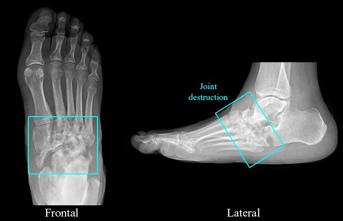
Neuropathic Arthritis / Charcot Foot

-There is preservation of bone mineral density.
-There is osseous fragmentation, midfoot fractures, and destruction of multiple tarsometatarsal joints.

Discussion
Neuropathic arthritis is caused by chronic repetitive trauma to insensitive joints. Underlying causes include diabetes, steroid use, trauma, alcoholism, multiple sclerosis, spina bifida, and syringomyelia. The radiologic findings are frequently discussed at the “6 D’s:” Density i.e. bone density maintained, Dislocation, Debris, Distention i.e. soft tissue swelling and joint effusions, Destruction, Disorganization.


http://eradiology.bidmc.harvard.edu/Classics/item.aspx?section=Emergency+Radiology&labelpk=33f0adab-853f-4010-a0e2-0756b3f1eac5&pk=001e927a-d1a5-417a-a4e1-954b594ef9ea

Lisfranc Fracture

-This patient demonstrates the medial cortical margin of the 2nd metatarsal base is malaligned with the medial margin of the middle cuneiform, termed a Lisfranc fracture.
-This is caused by severe plantar flexion of the foot.
-There are divergent and homolateral types.


http://eradiology.bidmc.harvard.edu/Classics/item.aspx?section=Emergency+Radiology&labelpk=33f0adab-853f-4010-a0e2-0756b3f1eac5&pk=d7301ffd-4d48-49f3-848e-c195254559b0

Jones Fracture

Findings
-This patient demonstrates a transverse fracture line at the base of the 5th metatarsal, called a Jones fracture.
-The fracture is located 1.5 – 2.5 cm from the proximal aspect of the 5th metatarsal.
-Typically caused by an inversion injury.
-Associated with a high incidence of non-union due to tenuous blood supply.


http://eradiology.bidmc.harvard.edu/Classics/item.aspx?section=Emergency+Radiology&labelpk=33f0adab-853f-4010-a0e2-0756b3f1eac5&pk=81e668ca-2c3a-4c55-bf1f-d67aeebd9ec0

Lover's Fracture


Findings

-This patient demonstrates a fracture through the calcaneus and a vertebral compression fracture at the thoracolumbar junction.

-This combination is called a “lover’s” fracture and is caused by axial loading (i.e. lover jumping from a window to escape an enraged spouse)

-There are retropulsed vertebral body fragments in the spinal canal, which can lead to neurologic compromise.

http://eradiology.bidmc.harvard.edu/Classics/item.aspx?section=Emergency+Radiology&labelpk=33f0adab-853f-4010-a0e2-0756b3f1eac5&pk=3e7aa04d-020a-401b-9bf2-f3558392e29b

Medullary Infarcts on Plain Films

Findings
-Medullary infarcts occur at the metaphyses.
-They are frequently multiple.
-They usually show well-defined dense geographic borders.
-They are frequently seen in patients on systemic steroids and sickle cell patients.


http://eradiology.bidmc.harvard.edu/Classics/item.aspx?section=Emergency+Radiology&labelpk=33f0adab-853f-4010-a0e2-0756b3f1eac5&pk=cbe389a4-219d-4de7-aaca-3acc9f52ddfd

Femoral Hernia with Small Bowel Obstruction on CT Scans


Findings

The CT scans demonstrate a bowel loop in the femoral canal with proximal small bowel dilatation.

Discussion

A femoral hernia lies in the femoral canal, medial to the femoral vein. Because of the narrowness of the femoral ring (the opening that forms the neck of a femoral hernia), it is more likely than an inguinal hernia to become incarcerated. Femoral hernias are most common in elderly women, typically presenting as a groin lump. The incidence of strangulation in femoral hernias is high.

http://eradiology.bidmc.harvard.edu/Classics/item.aspx?section=Emergency+Radiology&labelpk=33f0adab-853f-4010-a0e2-0756b3f1eac5&pk=431ef879-e8eb-4b25-85c2-ac1dfba5a398

Epidural Hemorrhage

Findings
The CT scans show a lens-shaped, biconvex high attenuation collection in the posterior left parietal lobe exerting mass effect.

Discussion
Acute bleeds present with high attenuation on CT. Epidural hematomas are due to traumatic head injuries which causes bleeding between the inner table of the skull and the dura. The majority are in the temporo-parietal region where a skull fracture causes damage to the middle meningeal artery or one of its branches. EDHs may cross the midline but do not cross suture lines.


http://eradiology.bidmc.harvard.edu/Classics/item.aspx?section=Emergency+Radiology&labelpk=33f0adab-853f-4010-a0e2-0756b3f1eac5&pk=c1634e61-5022-4bf4-8648-111c7af88cc3
Findings
The CT scans demonstrate a left frontal epidural hematoma.

Discussion
Epidural hematomas arise in the potential space between the dura and the skull and do not cross sutural margins; the bleeding is mostly due to rupture of the middle meningeal artery. The major cause for EDH is trauma and skull fractures are present in 75 to 95 percent of patients. Epidural bleeds produce a bi-convex (lens-shaped) hematoma on CT because the collection is limited by dural attachments at the cranial sutures.


http://eradiology.bidmc.harvard.edu/Classics/item.aspx?section=Emergency+Radiology&labelpk=33f0adab-853f-4010-a0e2-0756b3f1eac5&pk=e56ebea9-dcd1-44d4-9aea-bd6bfe3df54e

Misplaced ETT down Right Mainstem Bronchus on CXR

Findings
-The radiograph demonstrates the endotrachial tube in the right mainstem bronchus
-The right lung is hyperinflated with resultant flattening of the right hemidiaphragm.
-The left lung is collapsed with resultant elevation of the left hemidiaphragm and leftward shift of the mediastinum.


http://eradiology.bidmc.harvard.edu/Classics/item.aspx?section=Emergency+Radiology&labelpk=33f0adab-853f-4010-a0e2-0756b3f1eac5&pk=b0175468-097d-41ee-863e-d8471a453259

Fracture of the Radial Head with Effusion on Plain Films

Findings
-Frontal view: There is a subtle vertical fracture through the radial head.
-Lateral view: The anterior fat pad is displaced anteriorly. The posterior fat pad is visible. These indicate a joint effusion.

Discussion
The anterior fat pad is normally visualized hugging the anterior cortex of the distal humerus. The posterior fat pad is not seen on normal views. Anterior displacement of the anterior fat pad and visualization of the posterior fat pad are compatible with a joint effusion. Radial head fractures may be extremely difficult to detect on plain film. In the setting of acute trauma, if an elbow joint effusion is present, the presumption should be that a radial head fracture is present, even if the fracture line is not visible.


http://eradiology.bidmc.harvard.edu/Classics/item.aspx?section=Emergency+Radiology&labelpk=33f0adab-853f-4010-a0e2-0756b3f1eac5&pk=9cb9409c-877a-4252-87a7-b47e106a32b5

Comminuted Fracture of Distal Humerus

Findings
This patient presents with a comminuted fracture of the distal humerus from bullet shrapnel.

Discussion
A plate with screws was inplanted into this patient's arm for initial fixation of the fracture.


http://eradiology.bidmc.harvard.edu/Classics/item.aspx?section=Emergency+Radiology&labelpk=33f0adab-853f-4010-a0e2-0756b3f1eac5&pk=ecb9ac1b-483e-4e97-b8ab-71c57d883151