 Cortical destruction
-Cortical destruction is a common finding, and not very useful in distinguishing between malignant and benign lesions.
-Complete destruction may be seen in high-grade malignant lesions, but also in locally aggressive benign lesions like EG and osteomyelitis.
-More uniform cortical bone destruction can be found in benign and low-grade malignant lesions. -Endosteal scalloping of the cortical bone can be seen in benign lesions like FD and low-grade chondrosarcoma.The above images show irregular cortical destruction in an osteosarcoma (left) and cortical destruction with aggressive periosteal reaction in Ewing's sarcoma.
 - Ballooning is a special type of cortical destruction. In ballooning the destruction of endosteal cortical bone and the addition of new bone on the outside occur at the same rate, resulting in expansion.
-This 'neocortex' can be smooth and uninterrupted, but may also be focally interrupted in more aggressive lesions like GCT.
left: Chondromyxoid fibroma A benign, well-defined, expansile lesion with regular destruction of cortical bone and a peripheral layer of new bone.
right: Giant cell tumor A locally aggressive lesion with cortical destruction, expansion and a thin, interrupted peripheral layer of new bone. Notice the wide zone of transition towards the marrow cavity, which is a sign of aggressive behavior.
 Cortical destruction (3)
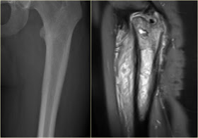
-In the group of malignant small round cell tumors which include Ewing's sarcoma, bone lymphoma and small cell osteosarcoma, the cortex may appear almost normal radiographically, while there is permeative growth throughout the Haversian channels.These tumors may be accompanied by a large soft tissue mass while there is almost no visible bone destruction.The image on the left shows an Ewing's sarcoma with permeative growth through the Haversian channels accompanied by a large soft tissue mass. The radiograph does not shown any signs of cortical destruction.
http://www.radiologyassistant.nl/en/494e15cbf0d8d
No comments:
Post a Comment