Best Bath Store
All Natural Blend of 14 Herbs Gentle Cleanse- $20 Off Colonix Now! Use Promo Code 20J
Amberen - Breakthrough Menopause Treatment
728x90
Custom Search

Thursday, May 20, 2010

multicystic water density structures(arrow) are noted peripherally connected to a dilated bile duct(open arrow).
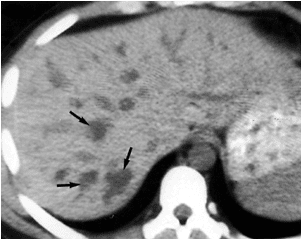
same as above.

T2 MRI image revealed hyper-intense cystic structure(arrow).


on T1MRI image ,it becomes hypo-intense.


The diagnosis now can be made with noninvasive imaging techniques including sonography, CT, and MRI. Cross-sectional imaging examinations show nonobstructive, saccular or beaded dilatation of the intrahepatic bile ducts. This is displayed as multiple, intrahepatic, rounded cystic spaces of varying sizes communicating with the branching, dilated, intrahepatic bile ducts .
One finding considered pathognomonic for Caroli disease is the central dot sign. This is characterized on unenhanced CT images by a small dot of slightly high attenuation in the dependent portion of the dilated bile duct. After the administration of intravenous contrast, these dots enhance intensely. The central dot sign is representative of portal venous radicles that are enveloped by, but not actually inside of, the markedly dilated bile ducts.
Reference:BiblioMed Textbook-Computed Body Tomography
showed bile dilatation in the left lobe and anterior segment of the right lobe with biloma(B) in the lateral segment of the left lobe.
left lobe bile duct dilatation with evidence of soft tissue mass(arrow) seen anterior to portal vein at the site of the common bile duct.

percutaneous trans hepatic cholangiogram showed
-straight arrows=dilated left bile ducts.
-curved arrow=abrupt cutoff seen proximal to junction between left and right bile ducts.


-G=multiple filling defects are seen in gall bladder suggestin calculi.


arrow=pneumobilia after PROCEDURE.
curved arrow=draining catheter.
m=mass.
Reference:BiblioMed Textbook-Computed Body Tomography


another case of cholangio-carcinoma


Reference:BiblioMed Textbook-Computed Body Tomography

CT signs:

ERCP showed irregular borders of bile ducts(arrow head)and the band like structures(straight arrows) of the intra hepatic ducts with the paucity of side branches corresponding to apperance
seen in A.
thickening of right and left hepatic ducts(arrows).
heterogenous enhancement of both lobes of the liver caused by cirrhosis or peri portal fibrosis.

(A)
open arrow=long segment of dilated duct without a normal branching pattern,this appearance of
cholangiographic pruned tree pattern. 1-In the evaluation of patients with PSC, CT has a role complementary to that of cholangiography. Computed tomography is not as sensitive as cholangiography for detecting the subtle mucosal abnormalities of the bile ducts, and therefore it should not be used as a screening examination in patients suspected of having PSC. However, the multiple, tight bile duct strictures in PSC can preclude adequate evaluation of the intrahepatic bile ducts with cholangiography; it is in this group of patients that CT can be helpful in evaluating the extent of disease.
2-The CT appearance characterised by presence of multiple areas of intra hepatic biliary dilatation in both lobes without any apparent connection to central ducts(skip dilatation).
Reference:BiblioMed Textbook-Computed Body Tomography


look to the image and describe
rhd,lhd=right and left hepatic biliary ducts are dilated centrally without evidence of peripheral dilatation.
black arrow=high density material found.
open arrow=gas in the bile duct.
AB=right peri hepatic abscess.

1-There is irregular, segmental dilatation of the intrahepatic bile ducts, with a predilection for the lateral segment of the left lobe and posterior segment of the right lobe. The dilatation primarily involves the larger central ducts, which taper and end abruptly. The peripheral ducts frequently are not visualized. The dilatation can be fusiform, varicose, or cavitary. The extrahepatic bile duct can be markedly dilated, even in the absence of dilatation of the intrahepatic ducts.


2-Intraductal stones can be detected by CT in 63% to 81% of patients. The stones can be in the intrahepatic ducts, in both the intra- and extrahepatic ducts, or only in the extrahepatic ducts.

Reference:BiblioMed Textbook-Computed Body Tomography
ascending cholangitis or acute cholangitis.


Bacterial infection of the biliary tree in the presence of complete or partial bile duct obstruction is called ascending cholangitis, acute cholangitis, bacterial cholangitis,

The CT findings that can be seen in patients with ascending cholangitis include
1-dilatation of the intra- and extrahepatic bile ducts,
2-increased attenuation of the bile because of the presence of intraductal debris,
3-diffuse and concentric bile duct wall thickening,
4-gas in the bile ducts or portal veins, and
5- intrahepatic abscesses (seen as low-attenuation areas in contiguity with the intrahepatic bile ducts).
MRI can show a similar spectrum of findings (Fig. ). Because bile duct dilatation can be absent in patients with acute or partial obstruction, there is no correlation between the presence and degree of bile duct dilatation and the severity of acute cholangitis. However, the presence of hepatic abscesses, pneumobilia, and portal venous gas are indicative of a poor prognosis.
Reference:BiblioMed Textbook-Computed Body Tomography

Wednesday, May 19, 2010

arrow=soft tissue density mass protruded
inside duodenum
Reference:BiblioMed Textbook-Computed Body Tomography

Reference:BiblioMed Textbook-Computed Body Tomography
chd=dilated common hepatic duct.
cbd=dilated common bile duct.
arrow=represent narrowed,displaced segment
due to extrinsic compression.
note,non visualisation of cystic duct and gall bladder.
Reference:BiblioMed Textbook-Computed Body Tomography
dense calcified mass(stone).
s=stent into common bile duct.

Reference:BiblioMed Textbook-Computed Body Tomography
crescent sign
s=soft tissue density calculus surrounded by
by a crescent area of water density bile
D=duodenum.
Reference:BiblioMed Textbook-Computed Body Tomography
Target sign
arrow=soft tissue density calculus surrounded by
a zone of water density bile(arrow head).
open arrow=dilated pancreatic duct in uncinate process.
Reference:BiblioMed Textbook-Computed Body Tomography
soft tissue density calculus
d=duodenum.
Reference:BiblioMed Textbook-Computed Body Tomography
arrow=CBD calculus.
p=pancreatic head.
d=duodenum.
Reference:BiblioMed Textbook-Computed Body Tomography

T1 image
T2 image


Reference:BiblioMed Textbook-Computed Body Tomography


multiple cystic areas seen in a branching pattern
similar to that of biliary pattern.
Reference:BiblioMed Textbook-Computed Body Tomography

Sunday, May 16, 2010

Reference:BiblioMed Textbook-Computed Body Tomography

Reference:BiblioMed Textbook-Computed Body Tomography

Reference:BiblioMed Textbook-Computed Body Tomography


Reference:BiblioMed Textbook-Computed Body Tomography

Reference:BiblioMed Textbook-Computed Body Tomography

Reference:BiblioMed Textbook-Computed Body Tomography

Reference:BiblioMed Textbook-Computed Body Tomography

Reference:BiblioMed Textbook-Computed Body Tomography