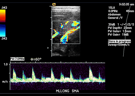
Normal spectral waveforms in the superior mesenteric artery. (a) Fasting waveform from a 26-day-old neonate shows a high-resistance flow pattern. (b) Postprandial waveform from a 6-day-old neonate shows a low-resistance pattern with an increase in diastolic flow velocity.
The celiac artery has a high-resistance pattern at its origin, with a small amount of reversed flow in early diastole. Its distal portion and branches lose the reversed early-diastolic flow component, showing continuous low-resistance forward flow throughout the cardiac cycle .
With the body in a fasting state, flow through the superior mesenteric artery has a high-resistance pattern, with a small amount of reversed flow in early diastole (Fig a). After a meal, the PSV and diastolic velocities increase. The reversed diastolic flow disappears, a low-resistance pattern develops, and the systolic spectral peak broadens (Fig b).

 The celiac artery arises from the anterior surface of the abdominal aorta and immediately divides into 3 branches: the common hepatic, the splenic and the left gastric arteries. The above ultrasound and Color Doppler images show the normal celiac artery with the 2 main branches, the splenic and common hepatic arteries. The left gastric is not visualized usually on sonography, being a small branch.