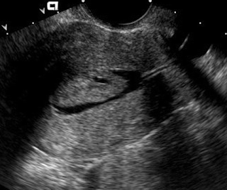
 Endometrial polyp. Sagittal sonohysterogram shows a large, well-defined mass in the fundus arising from the anterior aspect of the endometrium. Note the cystic area in the lower portion of the polyp.
http://www.google.com.eg/imgres?imgurl=http://radiographics.rsna.org/content/26/2/419/F12.small.gif&imgrefurl=http://radiographics.rsna.org/content/26/2/419.figures-only&usg=__VpUj1hDYgg3gkRJ5XSe6l8I_dXA=&h=192&w=200&sz=34&hl=en&start=44&itbs=1&tbnid=bQ3pvndRAhzw_M:&tbnh=100&tbnw=104&prev=/images%3Fq%3Dhysterosalpingography%26start%3D40%26hl%3Den%26sa%3DN%26gbv%3D2%26ndsp%3D20%26tbs%3Disch:1


No comments:
Post a Comment