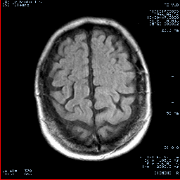
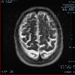
Figures 1, 2.
(1) Axial T2-weighted fast spin-echo
(2) Axial fast FLAIR (fluid-attenuated inversion recovery) image
Figure 3. Axial diffusion-weighted (z sensitizing direction) multishot echo-planar image (800/123)obtained with one signal acquisition and 6-mm
section thickness. The hyperintensity involving subcortical white matter and overlying cortex in the left parietal lobe is consistent with subacute infarction (arrow).
Acute Infarction
Early (within the first 6 hours after stroke) CT signs of brain ischemia are subtle and difficult to detect . On conventional MR images, early (within the first 6 hours after stroke) morphologic signs (produced by tissue swelling) are detected in 50% of acute infarctions; however, signal abnormalities are not detected . With diffusion-weighted imaging of acute infarction (within the first 6 hours after stroke), 94% sensitivity and 100% specificity have been reported ).
Early (within the first 6 hours after stroke) CT signs of brain ischemia are subtle and difficult to detect . On conventional MR images, early (within the first 6 hours after stroke) morphologic signs (produced by tissue swelling) are detected in 50% of acute infarctions; however, signal abnormalities are not detected . With diffusion-weighted imaging of acute infarction (within the first 6 hours after stroke), 94% sensitivity and 100% specificity have been reported ).
What's Going On?The interruption of cerebral blood flow results in rapid (within minutes) breakdown of energy metabolism and ion exchange pumps. This leads to a massive shift of water from the extracellular into the intracellular compartment (cytotoxic edema) and produces a typical high-intensity area on diffusion-weighted images.
Note! Diffusion-weighted imaging is the best technique for confirming a diagnosis of stroke in time to treat it with appropriate therapy. The value of diffusion-weighted and perfusion MR imaging in patient treatment decisions has not yet been clearly defined.


No comments:
Post a Comment