 A
A B
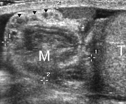
BTesticular torsion in a 12-year-old boy with right-sided scrotal pain of sudden onset. (a) Longitudinal US scan of the right hemiscrotum shows a round supratesticular mass (M), which represents an edematous spermatic cord. There are several anechoic structures (arrowheads) within the mass, which probably represent obstructed and dilated lymphatic vessels. T = testis. (b) Bilateral transverse color Doppler images show no color flow signals in the right testis, which is enlarged and has heterogeneous echogenicity. Reactive hydrocele (h) and thickening of the scrotal wall (*) are also seen. Testicular torsion and bell clapper deformity were confirmed at surgery.
No comments:
Post a Comment