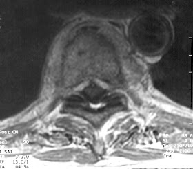


Clinical History: 76-year-old male with a history of back pain and suspicion of infection.
Radiologic Findings: T1-weighted images (Figs. 1 and 2) show, at the T8-9 level, decreased signal intensity in the disc with loss of delineation of the vertebral end plates from the disc. Mild central canal stenosis is noted. A T2-weighted image (Fig.3) demonstrates increased signal intensity in the intervertebral disc with loss of the normal intranuclear cleft of low signal in the center of the disc. Following Gadolinium administration, T1-weighted images (Figs. 4 and 5) show abnormal enhancement of the disc, adjacent vertebral bodies, and anterior epidural and paraspinal soft tissues.
Diagnosis: Discitis with adjacent osteomyelitis.
Discussion: Pyogenic vertebral discitis and osteomyelitis are related to bacterial seeding from hematogenous spread secondary to bacteremia, or extension from surgery, trauma, or from a contiguous infection. Hematogenous spread is the most common cause, primarily related to infections of the genitourinary tract, skin and respiratory tract.
The most common causative organisms are Staphylococcus aureus (60%) and Enterobacter species (30%). Tuberculous and fungal infections tend to spare the intervertebral disc until late in disease. The frequency of location, in decreasing order, is lumbar> thoracic > cervical spine.
Most cases respond to systemic antibiotics, although epidural abscesses may require surgical drainage, especially if there is cord compression.
The differential diagnosis of discitis and osteomyelitis includes degenerative disc disease, which usually shows decreased signal intensity in the disc on the T2-weighted images. Lymphoma and myeloma spare the disc. Postoperative discectomy may normally show enhancement of the disc space and posterior annulus, but enhancement of vertebral marrow is rare.
References: 1. Modic, et al., Radiologic Clinics of North America, Imaging of the Spine, W. B. Saunders.Philadelphia, PA, Vol. 29, No. 4, 1991, pgs. 813-821.
2. Willing, Atlas of Neuroradiology, W. B. Saunders, Philadelphia, PA, 1995, pgs. 545-548.
Submitted by:Rakesh Shah, M.D.Charles F. Lanzieri, M.D. (uhrad.com).
No comments:
Post a Comment