Best Bath Store
All Natural Blend of 14 Herbs Gentle Cleanse- $20 Off Colonix Now! Use Promo Code 20J
Amberen - Breakthrough Menopause Treatment
728x90
Custom Search

Saturday, May 15, 2010


Reference:BiblioMed Textbook-Computed Body Tomography


Reference:BiblioMed Textbook-Computed Body Tomography

Reference:BiblioMed Textbook-Computed Body Tomography

Reference:BiblioMed Textbook-Computed Body Tomography
this bronchogenic carcinoma showed extension of pulmonary
vessels inside it while in round atelectasis ,vessels deviate around
mass
Reference:BiblioMed Textbook-Computed Body Tomography

vessels passed around mass
Reference:BiblioMed Textbook-Computed Body Tomography
white arrows=dense calcified plaques.
black arrows=calcified mediastinal lymph nodes.
due to TB empyema.
Reference:BiblioMed Textbook-Computed Body Tomography
vertebral body 11 with bone destruction
and sclerosis
white arrow=thickened para spinal soft
tissue due inflammation.
Reference:BiblioMed Textbook-Computed Body Tomography
para-spinal pleural based lymphoma.
Reference:BiblioMed Textbook-Computed Body Tomography

pleural versus pulmonary lesion.
look to mild lung infiltrate surrounding
lesion denote that the lesion is of pulmonary
origin and not pleural.
Reference:BiblioMed Textbook-Computed Body Tomography

pleural nodule related to pleura covering right atrium.
Reference:BiblioMed Textbook-Computed Body Tomography
thin enhancing walls with obtuse angles with chest wall.
Reference:BiblioMed Textbook-Computed Body Tomography

1-spherical in shape.
2-thick irregular wall.
3-acute angle with chest wall.
4-related to a bronchus.
Reference:BiblioMed Textbook-Computed Body Tomography
E=lenticular shape with thin walls and smooth inner surface.
formin an obtuse angle with chest wall,pulmonary vessels are
compressed and displaced around empyema.
A=sherical in shape with thick iregular wall,and form acute
angle with chest wall,pulmonary vessels extend directly toward
abscess.
Reference:BiblioMed Textbook-Computed Body Tomography
lung abscess with acute angle with pleura.
it contains cavity with gas and fluid level.
Reference:BiblioMed Textbook-Computed Body Tomography
arrow=fistula between esophagus and empyema
in the right side(rupture esophagus).
Reference:BiblioMed Textbook-Computed Body Tomography
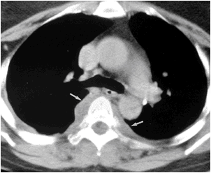
Em=empyema with thickened and enhanced
pleura.
Ef=no pleural thickening and enhancement
denoting effusion and not empyema.
presence of gas inside empyema suggest
broncho-pleural fistula.
open arrow=edema and inflammation of the
extra pleural tissue.
cephalad veiw showed empyema more rounded
simulating lung abscess.
Reference:BiblioMed Textbook-Computed Body Tomography

arrow head=thickened enhanced parietal pleura
arrow=thickening and inflammation of extra pleural
soft tissue in posterior hemithorax.
lung window setting showed bronchi and vessels
compressed and displaced by empyema.
Reference:BiblioMed Textbook-Computed Body Tomography

Friday, May 14, 2010

L=liver.
E=effusion.
A=ascites.
Reference:BiblioMed Textbook-Computed Body Tomography
E=pleural effusion.
arrow head=atelectatic lung.
A=ascites.
Reference:BiblioMed Textbook-Computed Body Tomography

E=effusion.
short arrow=bare area of liver.
long arrow=right crus.
Reference:BiblioMed Textbook-Computed Body Tomography
- thick black line represent right diaphragm
with its right crus.
-pleural effusion lies posterior and externally
to the diaphragm which may be extend behind
right crus adjacent to vertebra.
-ascites lies anterior and internal but does not
extend medially

Reference:BiblioMed Textbook-Computed Body Tomography
arrow head=bilateral small enhancing characteristic
pulmonary atelectasis.
bilateral small amount pleural effusion.
Reference:BiblioMed Textbook-Computed Body Tomography
H=heterogenous hyperdense pleural fluid causing
arrow=compressive collapse of the left lung.
Reference:BiblioMed Textbook-Computed Body Tomography
H=pleural hematoma.
Reference:BiblioMed Textbook-Computed Body Tomography
PL=loculated pleural effusion.
PC=pericardial effusion.
due to leukaemic infiltration.
Reference:BiblioMed Textbook-Computed Body Tomography
a-extra-pleural lesion=displace both parietal and visceral pleura
resulting in obtuse angle between lesion and chest wall.
+ or - rib erosion.
b-pleural lesion=arise between two layers of pleura producing obtuse
angle with chest wall or become pedunculated and protruded into peripheral
lung parenchyma producing acute angle with chest wall.
c-pulmonary lesion.
sub pleural pulmonary lesion cause acute angle with chest wall.
Reference:BiblioMed Textbook-Computed Body Tomography
arrow=inferior pulmonary ligament.
arrow head=thickening and enhancement
due to pleura due to radiation.
Reference:BiblioMed Textbook-Computed Body Tomography

CT section 2mm in thickness.
arrow head=lesser fissure(pausity of vascular markings).
arrow=greater fissure.