Best Bath Store
All Natural Blend of 14 Herbs Gentle Cleanse- $20 Off Colonix Now! Use Promo Code 20J
Amberen - Breakthrough Menopause Treatment
728x90
Custom Search

Saturday, May 1, 2010

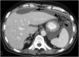
R=Right perihepatic space.
LA=left ant.perihepatic space.
LP=left post.perihepatic space.
LS=medial part of lesser sac.
arrowheads=gastroduodenal ligament.
m=metastases in falciform ligament.
AC=ascending colon.
DC=descending colon.
Small bowel mesentry.
RIS=right ilio-colic space
LIS=left ilio-colic space.
arrows indicate the nature flow of ascites.
arrowhead=hepatic serosal metastases.
M=malignant ascites in lesser sac.
L=lateral part of lesser sac
M=medial part.
arrowhead=left gastric artery passing through
a fold of peritonium.
mixture of fluid,air and oral contrast seen
in the right perihepatic peritoneal space.

A=perihepatic(subphrenic)abscess.
arrows=bare area of liver
abscess=fluid+air+fluid level+enhanced rim
or fluid+enhanced rim
arrow=fluid level in abscess.
arow head=ascites.
curved arrow=liver.
R=right perihepatic space.
SH=sub-hepatic space.
L=left sub-phrenic space.
LS=lesser sac.
PS=perisplenic space.
arrowhead=gastro-splenic ligament.
contrast and air in the anterior(a) and posterior(p) perihepatic spaces.

arrow=falciform ligament separate right and left peritoneal space.

metastatic nodule elevating external obligue muscle.
calcified metastases.
in a post-partum lady


abscess=fluid+air bubbles+enhancing rim+fluid level

contrast enhanced image showed a round
fluid collection in the left rectus abdominis muscle
arrow=extravasated contrast denote
active bleeding.

a defect between internal obligue and transversus muscle
arrowhead=external obligue muscle.
omental fat herniated through linea alba(arrowhead).
rm=rectus abdominis muscle.

Friday, April 30, 2010

adrenal pheochromocytoma( >3cm)

1-tumor of adrenal medulla.
2-produce excess catecholamine production.
3-when it arises outside adrenal gland , it is called para-ganglioma.
4-usually over 3cm when discovered.
5-homogenous soft tissue attenuation value with central necrosis.
6-heterogenous enhancement ,indistinguishable from adrenal neoplasm.

adrenal aldosteronism ( < 3cm)

1- due over-production of adrenal aldosterone.
2-they are usually less than 3cm,usually range between 5 to 35mm.
3-50% have attenuation similar to that near of water(-10 to +10HU).
-a low water attenuation lesion in patient with laboratory proven hyper-aldosteronism should not be diagnosed as an adrenal cyst.

D.D. of adrenal masses with attenuation similar to that of water:
1-aldosteronism.
2-adrenal metastases.
3-non functioning adenoma

adrenal cushing syndrome( >3cm)

1-etiology:excess production of cortison by adrenal cortex,due to excess production of ACTH,resulting from adrenal adenoma or hyperplasia.
2-CT adrenal is done in case of cushing disease to distinguish ACTH dependent(hyperplasia) from independent type(focal mass disorders) and to determined the site of this focal mass disorders.
3-CT findings of adenoma
-size:more than 2cm,from 2 to 5 cm in diameter.
-smooth,round or oval and homogenous with little enhancement.
-mostly are of soft tissue attenuation,but it could be near water attenuation(relative high fat content).
4-it should be noted that the CT appearance of different types of adenomas are indistinguishable whether they produce excess cortisol or aldesterone or they are not hyperfunctioning.

adrenal hemorrhage

hyperdense adrenal lesion

D.D. of bilateral adrenal masses:
A)with cystic density:
1-simple adrenal cyst
2-complicated cyst such as
-adrenal metastases.
-calcified adrenal cyst.
3-histoplasmosis.
4-TB - or + calcification
5-non functioning adrenal adenoma.
B)with soft tissue density:
-adrenal metastases.
-lymphoma.
-pheochromocytoma in MEN 2 b(central necrosis).
C)with fat density:
-myelolipoma - or + hemorrhage.
D)blood density:
1-adrenal hemorrhage.

bilateral adrenal histoplasmosis


adrenal tuberculosis


adrenal cyst

left adrenal cyst with thick calcification.

bilateral adrenal myelolipoma with hemorrhage


adrenal myelolipoma


adrenal lymphoma


adrenal metastases


non functioning adenoma

1-homogenous near water attenuation
ch.ch.of non functioning adenoma.
2-low attenuation of adrenal mass
exclude malignancy.

pheochromocytoma

D.D.from adrenal carcinoma.
(by laboratory findings).
D.D. of heterogenous enhancement of adrenal masses.
-pheochromocytoma.
-adrenal carcinoma

multiple pheochromocytomas in MEN2b


pheochromocytoma

right adrenal mass


1-contrast enhancing CT.
2-Low attenuation mass
with irregular enhancing
nodules peripherally.

aldosteronomas


cushing syndrome


smooth thickening of both adrenals.

Thursday, April 29, 2010

right perinephric abscess.
left angiomyolipoma with hemorrhage.


high level of creatinine.

persistent dense nephrogram
with opacification of gall
bladder.
calcification limited to medullary pyramids.

1-bilateral swollen kidney.
2-poorly nhancing pyramids.